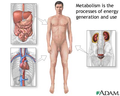
Metabolism


Metabolism Basics

Specific proteins in the body control the chemical reactions of metabolism, and each chemical reaction is coordinated with other body functions. In fact, thousands of metabolic reactions happen at the same time — all regulated by the body — to keep our cells healthy and working.

Metabolism is a constant process that begins when we're conceived and ends when we die. It is a vital process for all life forms — not just humans. If metabolism stops, a living thing dies.

Here's an example of how the process of metabolism works in humans — and it begins with plants: First, a green plant takes in energy from sunlight. The plant uses this energy and the molecule cholorophyll (which gives plants their green color) to build sugars from water and carbon dioxide in a process known as photosynthesis.

When people and animals eat the plants (or, if they're carnivores, when they eat animals that have eaten the plants), they take in this energy (in the form of sugar), along with other vital cell-building chemicals. The body's next step is to break the sugar down so that the energy released can be distributed to, and used as fuel by, the body's cells.

After food is eaten, molecules in the digestive system called enzymes break proteins down into amino acids, fats into fatty acids, and carbohydrates into simple sugars (e.g., glucose). In addition to sugar, both amino acids and fatty acids can be used as energy sources by the body when needed. These compounds are absorbed into the blood, which transports them to the cells.

After they enter the cells, other enzymes act to speed up or regulate the chemical reactions involved with "metabolizing" these compounds. During these processes, the energy from these compounds can be released for use by the body or stored in body tissues, especially the liver, muscles, and body fat.

In this way, the process of metabolism is really a balancing act involving two kinds of activities that go on at the same time — the building up of body tissues and energy stores and the breaking down of body tissues and energy stores to generate more fuel for body functions.
  • Anabolism, or constructive metabolism, is all about building and storing: It supports the growth of new cells, the maintenance of body tissues, and the storage of energy for use in the future. During anabolism, small molecules are changed into larger, more complex molecules of carbohydrate, protein, and fat.
  • Catabolism, or destructive metabolism, is the process that produces the energy required for all activity in the cells. In this process, cells break down large molecules (mostly carbohydrates and fats) to release energy. This energy release provides fuel for anabolism, heats the body, and enables the muscles to contract and the body to move. As complex chemical units are broken down into more simple substances, the waste products released in the process of catabolism are removed from the body through the skin, kidneys, lungs, and intestines.

Several of the hormones of the endocrine system are involved in controlling the rate and direction of metabolism. Thyroxine, a hormone produced and released by the thyroid gland, plays a key role in determining how fast or slow the chemical reactions of metabolism proceed in a person's body.

Another gland, the pancreas secretes hormones that help determine whether the body's main metabolic activity at a particular time will be anabolic or catabolic. For example, after eating a meal, usually more anabolic activity occurs because eating increases the level of glucose — the body's most important fuel — in the blood. The pancreas senses this increased level of glucose and releases the hormone insulin, which signals cells to increase their anabolic activities.

Metabolism is a complicated chemical process, so it's not surprising that many people think of it in its simplest sense: as something that influences how easily our bodies gain or lose weight. That's where calories come in. A calorie is a unit that measures how much energy a particular food provides to the body. A chocolate bar has more calories than an apple, so it provides the body with more energy — and sometimes that can be too much of a good thing. Just as a car stores gas in the gas tank until it is needed to fuel the engine, the body stores calories — primarily as fat. If you overfill a car's gas tank, it spills over onto the pavement. Likewise, if a person eats too many calories, they "spill over" in the form of excess body fat.

The number of calories someone burns in a day is affected by how much that person exercises, the amount of fat and muscle in his or her body, and the person's basal metabolic rate (or BMR). BMR is a measure of the rate at which a person's body "burns" energy, in the form of calories, while at rest. The BMR can play a role in someone's tendency to gain weight. For example, a person with a low BMR (who therefore burns fewer calories while at rest or sleeping) will tend to gain more pounds of body fat over time, compared with a similar-sized person with an average BMR who eats the same amount of food and gets the same amount of exercise.

What factors influence BMR? To a certain extent, BMR is inherited. Sometimes health problems can affect BMR. But people can actually change their BMR in certain ways. For example, exercising more will not only cause a person to burn more calories directly from the extra activity itself, but becoming more physically fit will increase BMR as well. BMR is also influenced by body composition — people with more muscle and less fat generally have higher BMRs.

Lungs and Respiratory System


About the Lungs and Respiratory System
At the top of the respiratory system, the nostrils (also called nares) act as the air intake, bringing air into the nose, where it's warmed and humidified. Tiny hairs called cilia protect the nasal passageways and other parts of the respiratory tract, filtering out dust and other particles that enter the nose through the breathed air.

Air can also be taken in through the mouth. These two openings of the airway (the nasal cavity and the mouth) meet at the pharynx, or throat, at the back of the nose and mouth. The pharynx is part of the digestive system as well as the respiratory system because it carries both food and air. At the bottom of the pharynx, this pathway divides in two, one for food (the esophagus, which leads to the stomach) and the other for air. The epiglottis, a small flap of tissue, covers the air-only passage when you swallow, keeping food and liquid from going into the lungs.

The larynx, or voice box, is the uppermost part of the air-only pipe. This short tube contains a pair of vocal cords, which vibrate to make sounds. The trachea, or windpipe, extends downward from the base of the larynx. It lies partly in the neck and partly in the chest cavity. The walls of the trachea are strengthened by stiff rings of cartilage to keep it open. The trachea is also lined with cilia, which sweep fluids and foreign particles out of the airway so that they stay out of the lungs.

At its bottom end, the trachea divides into left and right air tubes called bronchi, which connect to the lungs. Within the lungs, the bronchi branch into smaller bronchi and even smaller tubes called bronchioles. Bronchioles end in tiny air sacs called alveoli, where the exchange of oxygen and carbon dioxide actually takes place. Each lung houses about 300 to 400 million alveoli. The lungs also contain elastic tissues that allow them to inflate and deflate without losing shape and are encased by a thin lining called the pleura. This network of alveoli, bronchioles, and bronchi is known as the bronchial tree.

The chest cavity, or thorax, is the airtight box that houses the bronchial tree, lungs, heart, and other structures. The top and sides of the thorax are formed by the ribs and attached muscles, and the bottom is formed by a large muscle called the diaphragm. The chest walls form a protective cage around the lungs and other contents of the chest cavity. Separating the chest from the abdomen, the diaphragm plays a lead role in breathing. It moves downward when we breathe in, enlarging the chest cavity and pulling air in through the nose or mouth. When we breathe out, the diaphragm moves upward, forcing the chest cavity to get smaller and pushing the gases in the lungs up and out of the nose and mouth.

What the Lungs and Respiratory System Do
The air we breathe is made up of several gases. Oxygen is the most important for keeping us alive because body cells need it for energy and growth. Without oxygen, the body's cells would die.

Carbon dioxide is the waste gas produced when carbon is combined with oxygen as part of the energy-making processes of the body. The lungs and respiratory system allow oxygen in the air to be taken into the body, while also enabling the body to get rid of carbon dioxide in the air breathed out.

Respiration is the set of events that results in the exchange of oxygen from the environment and carbon dioxide from the body's cells. The process of taking air into the lungs is inspiration, or inhalation, and the process of breathing it out is expiration, or exhalation.

Air is inhaled through the mouth or through the nose. Cilia lining the nose and other parts of the upper respiratory tract move back and forth, pushing foreign matter that comes in with air (like dust) either toward the nostrils to be expelled or toward the pharynx. The pharynx passes the foreign matter along to the stomach to eventually be eliminated by the body. As air is inhaled, the mucous membranes of the nose and mouth warm and humidify the air before it enters the lungs.

When you breathe in, the diaphragm moves downward toward the abdomen, and the rib muscles pull the ribs upward and outward. In this way, the volume of the chest cavity is increased. Air pressure in the chest cavity and lungs is reduced, and because gas flows from high pressure to low, air from the environment flows through the nose or mouth into the lungs. In exhalation, the diaphragm moves upward and the chest wall muscles relax, causing the chest cavity to contract. Air pressure in the lungs rises, so air flows from the lungs and up and out of respiratory system through the nose or mouth.

Every few seconds, with each inhalation, air fills a large portion of the millions of alveoli. In a process called diffusion, oxygen moves from the alveoli to the blood through the capillaries (tiny blood vessels) lining the alveolar walls. Once in the bloodstream, oxygen gets picked up by the hemoglobin in red blood cells. This oxygen-rich blood then flows back to the heart, which pumps it through the arteries to oxygen-hungry tissues throughout the body. In the tiny capillaries of the body tissues, oxygen is freed from the hemoglobin and moves into the cells. Carbon dioxide, which is produced during the process of diffusion, moves out of these cells into the capillaries, where most of it is dissolved in the plasma of the blood. Blood rich in carbon dioxide then returns to the heart via the veins. From the heart, this blood is pumped to the lungs, where carbon dioxide passes into the alveoli to be exhaled.

Kidneys and Urinary Tract


Our bodies produce several kinds of wastes, including sweat, carbon dioxide gas, feces (stool), and urine. These wastes exit the body in different ways. Sweat is released through pores in the skin. Water vapor and carbon dioxide are exhaled from the lungs. And undigested food materials are formed into feces in the intestines and excreted from the body as solid waste in bowel movements.

Urine, which is produced by the kidneys, contains the byproducts of metabolism — salts, toxins, and water — that end up in the blood. The kidneys and urinary tract (which includes the kidneys, ureters, bladder, and urethra) filter and eliminate these waste substances from our blood. Without the kidneys, waste products and toxins would soon build up in the blood to dangerous levels.

In addition to eliminating wastes, the kidneys and urinary tract also regulate many important body functions. For example, the kidneys monitor and maintain the body's balance of water, ensuring that our tissues receive enough water to function properly and be healthy.

When doctors take a urine sample, the results reveal how well the kidneys are working. For example, blood, protein, or white blood cells in the urine may indicate injury, inflammation, or infection of the kidneys, and glucose in the urine may be an indication of diabetes.

What the Kidneys and Urinary Tract Do

Although the two kidneys work together to perform many vital functions, people can live a normal, healthy life with just one kidney. In fact, some people are born with just one of these bean-shaped organs. If one kidney is removed, the remaining one will enlarge within a few months to take over the role of filtering blood on its own.

Every minute, more than 1 quart (about 1 liter) of blood goes to the kidneys. About one fifth of the blood pumped from the heart goes to the kidneys at any one time.

In addition to filtering blood, producing urine, and ensuring that body tissues receive enough water, the kidneys also regulate blood pressure and the level of vital salts in the blood. By regulating salt levels through production of an enzyme called renin (as well as other substances), the kidneys ensure that blood pressure is regulated.

The kidneys also secrete the hormone erythropoietin, which stimulates and controls red blood cell production (red blood cells carry oxygen throughout the body). In addition, the kidneys help regulate the acid-base balance (or the pH) of the blood and body fluids, which is necessary for the body to function normally.

How the Kidneys and Urinary Tract Work

The kidneys are located just under the ribcage in the back, one on each side. The right kidney is located below the liver, so it's a little lower than the left one. Each adult kidney is about the size of a fist. Each has an outer layer called the cortex, which contains the filtering units.

The center part of the kidney, the medulla has 10 to 15 fan-shaped structures called pyramids. These drain urine into cup-shaped tubes called calyxes. A layer of fat surrounds the kidneys to cushion and help hold them in place.

Here's how the kidneys filter blood : Blood travels to each kidney through the renal artery, which enters the kidney at the hilus, the indentation in the kidney that gives it its bean shape. As it enters the cortex, the artery branches to envelope the nephrons — 1 million tiny filtering units in each kidney that remove the harmful substances from the blood.

Each of the nephrons contain a filter called the glomerulus, which contains a network of tiny blood vessels known as capillaries. The fluid filtered from the blood by the glomerulus then travels down a tiny tube-like structure called a tubule, which adjusts the level of salts, water, and wastes that are excreted in the urine.

Filtered blood leaves the kidney through the renal vein and flows back to the heart.

The continuous blood supply entering and leaving the kidneys gives the kidneys their dark red color. While the blood is in the kidneys, water and some of the other blood components (such as acids, glucose, and other nutrients) are reabsorbed back into the bloodstream. Left behind is urine. Urine is a concentrated solution of waste material containing water, urea, a waste product that forms when proteins are broken down), salts, amino acids, byproducts of bile from the liver, ammonia, and any substances that cannot be reabsorbed into the blood. Urine also contains urochrome, a pigmented blood product that gives urine its yellowish color.

The renal pelvis, located near the hilus, collects the urine flowing from the calyxes. From the renal pelvis, urine is transported out of the kidneys through the ureters, tubes that carry the urine out of each kidney to be stored in the urinary bladder — a muscular collection sac in the lower abdomen.

The bladder expands as it fills and can hold about 2 cups (half a liter) of urine at any given time (an average adult produces about 6 cups, or 1½ liters, of urine per day). An adult needs to produce and excrete at least one third of this amount in order to adequately clear waste products from the body. Producing too much or not enough urine may indicate illness.

When the bladder is full, nerve endings in its wall send impulses to the brain. When a person is ready to urinate, the bladder walls contract and the sphincter (a ring-like muscle that guards the exit from the bladder to the urethra) relaxes. The urine is ejected from the bladder and out of the body through the urethra, another tube-like structure. The male urethra ends at the tip of the penis; the female urethra ends just above the vaginal opening.

Immune System


The immune system, which is made up of special cells, proteins, tissues, and organs, defends people against germs and microorganisms every day. In most cases, the immune system does a great job of keeping people healthy and preventing infections. But sometimes problems with the immune system can lead to illness and infection.

What the Immune System Does

The immune system is the body's defense against infectious organisms and other invaders. Through a series of steps called the immune response, the immune system attacks organisms and substances that invade our systems and cause disease. The immune system is made up of a network of cells, tissues, and organs that work together to protect the body.

The cells that are part of this defense system are white blood cells, or leukocytes. They come in two basic types (more on these below), which combine to seek out and destroy the organisms or substances that cause disease.

Leukocytes are produced or stored in many locations throughout the body, including the thymus, spleen, and bone marrow. For this reason, they are called the lymphoid organs. There are also clumps of lymphoid tissue throughout the body, primarily in the form of lymph nodes, that house the leukocytes.

The leukocytes circulate through the body between the organs and nodes by means of the lymphatic vessels. Leukocytes can also circulate through the blood vessels. In this way, the immune system works in a coordinated manner to monitor the body for germs or substances that might cause problems.

The two basic types of leukocytes are:
  • phagocytes, cells that chew up invading organisms
  • lymphocytes, cells that allow the body to remember and recognize previous invaders and help the body destroy them

A number of different cells are considered phagocytes. The most common type is the neutrophil, which primarily fights bacteria. If doctors are worried about a bacterial infection, they might order a blood test to see if a patient has an increased number of neutrophils triggered by the infection. Other types of phagocytes have their own jobs to make sure that the body responds appropriately to a specific type of invader.

There are two kinds of lymphocytes: the B lymphocytes and the T lymphocytes. Lymphocytes start out in the bone marrow and either stay there and mature into B cells, or they leave for the thymus gland, where they mature into T cells. B lymphocytes and T lymphocytes have separate jobs to do: B lymphocytes are like the body's military intelligence system, seeking out their targets and sending defenses to lock onto them. T cells are like the soldiers, destroying the invaders that the intelligence system has identified. Here's how it works.

Antigens are foreign substances that invade the body. When an antigen is detected, several types of cells work together to recognize and respond to it. These cells trigger the B lymphocytes to produce antibodies, specialized proteins that lock onto specific antigens. Antibodies and antigens fit together like a key and a lock.

Once the B lymphocytes have produced antibodies, these antibodies continue to exist in a person's body, so that if the same antigen is presented to the immune system again, the antibodies are already there to do their job. That's why if someone gets sick with a certain disease, like chickenpox, that person typically doesn't get sick from it again. This is also why we use immunizations to prevent getting certain diseases. The immunization introduces the body to the antigen in a way that doesn't make a person sick, but it does allow the body to produce antibodies that will then protect that person from future attack by the germ or substance that produces that particular disease.

Although antibodies can recognize an antigen and lock onto it, they are not capable of destroying it without help. That is the job of the T cells. The T cells are part of the system that destroys antigens that have been tagged by antibodies or cells that have been infected or somehow changed. (There are actually T cells that are called "killer cells.") T cells are also involved in helping signal other cells (like phagocytes) to do their jobs.

Antibodies can also neutralize toxins (poisonous or damaging substances) produced by different organisms. Lastly, antibodies can activate a group of proteins called complement that are also part of the immune system. Complement assists in killing bacteria, viruses, or infected cells.

All of these specialized cells and parts of the immune system offer the body protection against disease. This protection is called immunity.

Humans have three types of immunity — innate, adaptive, and passive:

Innate Immunity

Everyone is born with innate (or natural) immunity, a type of general protection that humans have. Many of the germs that affect other species don't harm us. For example, the viruses that cause leukemia in cats or distemper in dogs don't affect humans. Innate immunity works both ways because some viruses that make humans ill — such as the virus that causes HIV/AIDS — don't make cats or dogs sick either.

Innate immunity also includes the external barriers of the body, like the skin and mucous membranes (like those that line the nose, throat, and gastrointestinal tract), which are our first line of defense in preventing diseases from entering the body. If this outer defensive wall is broken (like if you get a cut), the skin attempts to heal the break quickly and special immune cells on the skin attack invading germs.

Adaptive Immunity

We also have a second kind of protection called adaptive (or active) immunity. This type of immunity develops throughout our lives. Adaptive immunity involves the lymphocytes (as in the process described above) and develops as children and adults are exposed to diseases or immunized against diseases through vaccination.

Passive Immunity

Passive immunity is "borrowed" from another source and it lasts for a short time. For example, antibodies in a mother's breast milk provide an infant with temporary immunity to diseases that the mother has been exposed to. This can help protect the infant against infection during the early years of childhood.

Everyone's immune system is different. Some people never seem to get infections, whereas others seem to be sick all the time. As people get older, they usually become immune to more germs as the immune system comes into contact with more and more of them. That's why adults and teens tend to get fewer colds than kids — their bodies have learned to recognize and immediately attack many of the viruses that cause colds.

Heart and Circulatory System


About the Heart and Circulatory System

The circulatory system is composed of the heart and blood vessels, including arteries, veins, and capillaries. Our bodies actually have two circulatory systems: The pulmonary circulation is a short loop from the heart to the lungs and back again, and the systemic circulation (the system we usually think of as our circulatory system) sends blood from the heart to all the other parts of our bodies and back again.

The heart is the key organ in the circulatory system. As a hollow, muscular pump, its main function is to propel blood throughout the body. It usually beats from 60 to 100 times per minute, but can go much faster when necessary. It beats about 100,000 times a day, more than 30 million times per year, and about 2.5 billion times in a 70-year lifetime.

The heart gets messages from the body that tell it when to pump more or less blood depending on an individual's needs. When we're sleeping, it pumps just enough to provide for the lower amounts of oxygen needed by our bodies at rest. When we're exercising or frightened, the heart pumps faster to increase the delivery of oxygen.

The heart has four chambers that are enclosed by thick, muscular walls. It lies between the lungs and just to the left of the middle of the chest cavity. The bottom part of the heart is divided into two chambers called the right and left ventricles, which pump blood out of the heart. A wall called the interventricular septum divides the ventricles.

The upper part of the heart is made up of the other two chambers of the heart, the right and left atria. The right and left atria receive the blood entering the heart. A wall called the interatrial septum divides the right and left atria, which are separated from the ventricles by the atrioventricular valves. The tricuspid valve separates the right atrium from the right ventricle, and the mitral valve separates the left atrium and the left ventricle.

Two other cardiac valves separate the ventricles and the large blood vessels that carry blood leaving the heart. These are the pulmonic valve, which separates the right ventricle from the pulmonary artery leading to the lungs, and the aortic valve, which separates the left ventricle from the aorta, the body's largest blood vessel.

Arteries carry blood away from the heart. They are the thickest blood vessels, with muscular walls that contract to keep the blood moving away from the heart and through the body. In the systemic circulation, oxygen-rich blood is pumped from the heart into the aorta. This huge artery curves up and back from the left ventricle, then heads down in front of the spinal column into the abdomen. Two coronary arteries branch off at the beginning of the aorta and divide into a network of smaller arteries that provide oxygen and nourishment to the muscles of the heart.

Unlike the aorta, the body's other main artery, the pulmonary artery, carries oxygen-poor blood. From the right ventricle, the pulmonary artery divides into right and left branches, on the way to the lungs where blood picks up oxygen.

Arterial walls have three layers:
  1. The endothelium is on the inside and provides a smooth lining for blood to flow over as it moves through the artery.
  2. The media is the middle part of the artery, made up of a layer of muscle and elastic tissue.
  3. The adventitia is the tough covering that protects the outside of the artery.

As they get farther from the heart, the arteries branch out into arterioles, which are smaller and less elastic.

Veins carry blood back to the heart. They're not as muscular as arteries, but they contain valves that prevent blood from flowing backward. Veins have the same three layers that arteries do, but are thinner and less flexible. The two largest veins are the superior and inferior vena cavae. The terms superior and inferior don't mean that one vein is better than the other, but that they're located above and below the heart.

A network of tiny capillaries connects the arteries and veins. Though tiny, the capillaries are one of the most important parts of the circulatory system because it's through them that nutrients and oxygen are delivered to the cells. In addition, waste products such as carbon dioxide are also removed by the capillaries.

What the Heart and Circulatory System Do

The circulatory system works closely with other systems in our bodies. It supplies oxygen and nutrients to our bodies by working with the respiratory system. At the same time, the circulatory system helps carry waste and carbon dioxide out of the body.

Hormones — produced by the endocrine system — are also transported through the blood in the circulatory system. As the body's chemical messengers, hormones transfer information and instructions from one set of cells to another. For example, one of the hormones produced by the heart helps control the kidneys' release of salt from the body.

One complete heartbeat makes up a cardiac cycle, which consists of two phases:
  1. In the first phase, the ventricles contract (this is called systole), sending blood into the pulmonary and systemic circulation. To prevent the flow of blood backwards into the atria during systole, the atrioventricular valves close, creating the first sound (the lub). When the ventricles finish contracting, the aortic and pulmonary valves close to prevent blood from flowing back into the ventricles. This is what creates the second sound (the dub).
  2. Then the ventricles relax (this is called diastole) and fill with blood from the atria, which makes up the second phase of the cardiac cycle.

A unique electrical conduction system in the heart causes it to beat in its regular rhythm. The sinoatrial or SA node, a small area of tissue in the wall of the right atrium, sends out an electrical signal to start the contracting of the heart muscle. This node is called the pacemaker of the heart because it sets the rate of the heartbeat and causes the rest of the heart to contract in its rhythm.

These electrical impulses cause the atria to contract first, and then travel down to the atrioventricular or AV node, which acts as a kind of relay station. From here the electrical signal travels through the right and left ventricles, causing them to contract and forcing blood out into the major arteries.

In the systemic circulation, blood travels out of the left ventricle, to the aorta, to every organ and tissue in the body, and then back to the right atrium. The arteries, capillaries, and veins of the systemic circulatory system are the channels through which this long journey takes place.

Once in the arteries, blood flows to smaller arterioles and then to capillaries. While in the capillaries, the bloodstream delivers oxygen and nutrients to the body's cells and picks up waste materials. Blood then goes back through the capillaries into venules, and then to larger veins until it reaches the vena cavae.

Blood from the head and arms returns to the heart through the superior vena cava, and blood from the lower parts of the body returns through the inferior vena cava. Both vena cavae deliver this oxygen-depleted blood into the right atrium. From here the blood exits to fill the right ventricle, ready to be pumped into the pulmonary circulation for more oxygen.

In the pulmonary circulation, blood low in oxygen but high in carbon dioxide is pumped out the right ventricle into the pulmonary artery, which branches off in two directions. The right branch goes to the right lung, and vice versa.

In the lungs, the branches divide further into capillaries. Blood flows more slowly through these tiny vessels, allowing time for gases to be exchanged between the capillary walls and the millions of alveoli, the tiny air sacs in the lungs.

During the process called oxygenation, oxygen is taken up by the bloodstream. Oxygen locks onto a molecule called hemoglobin in the red blood cells. The newly oxygenated blood leaves the lungs through the pulmonary veins and heads back to the heart. It enters the heart in the left atrium, then fills the left ventricle so it can be pumped into the systemic circulation.