Here's how each aspect of the mouth and teeth plays an important role in our daily lives.
Basic Anatomy of the Mouth and Teeth
The entrance to the digestive tract, the mouth is lined with mucous membranes. The membrane-covered roof of the mouth is called the palate. The front part consists of a bony portion called the hard palate, with a fleshy rear part called the soft palate. The hard palate divides the mouth and the nasal passages above. The soft palate forms a curtain between the mouth and the throat, or pharynx, to the rear. The soft palate contains the uvula, the dangling flesh at the back of the mouth. The tonsils are located on either side of the uvula and look like twin pillars holding up the opening to the pharynx.
A bundle of muscles extends from the floor of the mouth to form the tongue. The upper surface of the tongue is covered with tiny bumps called papillae. These contain tiny pores that are our taste buds. Four kinds of taste buds are grouped together on certain areas of the tongue — those that sense sweet, salty, sour, and bitter tastes. Three pairs of salivary glands secrete saliva, which contains a digestive enzyme called amylase that starts the breakdown of carbohydrates even before food enters the stomach.
The lips are covered with skin on the outside and with slippery mucous membranes on the inside of the mouth. The major lip muscle, called the orbicularis oris, allows for the lips' mobility. The reddish tint of the lips comes from underlying blood vessels. The inside portion of both lips is connected to the gums.
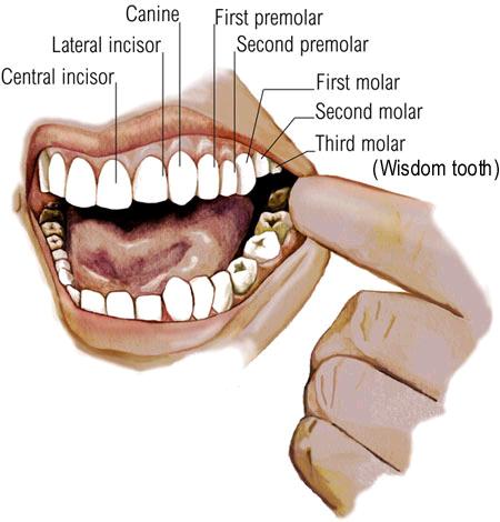
There are several types of teeth. Incisors are the squarish, sharp-edged teeth in the front of the mouth. There are four on the bottom and four on the top. On either side of the incisors are the sharp canines. The upper canines are sometimes called eyeteeth. Behind the canines are the premolars, or bicuspids. There are two sets, or four premolars, in each jaw.
The molars, situated behind the premolars, have points and grooves. There are 12 molars — three sets in each jaw called the first, second, and third molars. The third molars are the wisdom teeth, thought by some to have evolved thousands of years ago when human diets consisted of mostly raw foods that required extra chewing power. But because they can crowd out the other teeth, sometimes a dentist will need to remove them.
Human teeth are made up of four different types of tissue: pulp, dentin, enamel, and cementum. The pulp is the innermost portion of the tooth and consists of connective tissue, nerves, and blood vessels, which nourish the tooth. The pulp has two parts — the pulp chamber, which lies in the crown, and the root canal, which is in the root of the tooth. Blood vessels and nerves enter the root through a small hole in its tip and extend through the canal into the pulp chamber.
Dentin surrounds the pulp. A hard yellow substance consisting mostly of mineral salts and water, it makes up most of the tooth and is as hard as bone. It's the dentin that gives teeth their yellowish tint. Enamel, the hardest tissue in the body, covers the dentin and forms the outermost layer of the crown. It enables the tooth to withstand the pressure of chewing and protects it from harmful bacteria and changes in temperature from hot and cold foods. Both the dentin and pulp extend into the root. A bony layer of cementum covers the outside of the root, under the gum line, and holds the tooth in place within the jawbone. Cementum is also as hard as bone.
Normal Development of the Mouth and Teeth
Humans are diphyodont, meaning that they develop two sets of teeth. The first set of 20 deciduous teeth are also called the milk, primary, temporary, falling-off, or baby teeth. They begin to develop before birth and begin to fall out when a child is around 6 years old. They're replaced by a set of 32 permanent teeth, which are also called secondary or adult teeth.
Around the 8th week after conception, oval-shaped tooth buds consisting of cells form in the embryo. These buds begin to harden about the 16th week. Although teeth aren't visible at birth, both the primary and permanent teeth are forming below the gums. The crown, or the hard enamel-covered part that's visible in the mouth, develops first. When the crown is fully grown, the root begins to develop.
Between the ages of 6 months and 1 year, the deciduous teeth begin to push through the gums. This process is called eruption or teething. At this point, the crown is complete and the root is almost fully formed. By the time a child is 3 years old, he or she has a set of 20 deciduous teeth, 10 in the lower and 10 in the upper jaw. Each jaw has four incisors, two canines, and four molars. The molars' purpose is to grind food, and the incisors and canine teeth are used to bite into and tear food.
The primary teeth help the permanent teeth erupt in their normal positions; most of the permanent teeth form close to the roots of the primary teeth. When a primary tooth is preparing to fall out, its root begins to dissolve. This root has completely dissolved by the time the permanent tooth below it is ready to erupt.
Kids start to lose their primary teeth, or baby teeth, at about 6 years old. This begins a phase of permanent tooth development that lasts over the next 15 years, as the jaw steadily grows into its adult form. From ages 6 to 9, the incisors and first molars start to come in. Between ages 10 and 12, the first and second premolars, as well as the canines, erupt. From 12 to 13, the second molars come in. The wisdom teeth (third molars) erupt between the ages of 17 and 21.
Sometimes there isn't room in a person's mouth for all the permanent teeth. If this happens, the wisdom teeth may not come through at all. Overcrowding of the teeth is one of the reasons kids get braces.
What the Mouth and Teeth Do
The first step of digestion involves the mouth and teeth. Food enters the mouth and is immediately broken down into smaller pieces by our teeth. Each type of tooth serves a different function in the chewing process. Incisors cut foods when you bite into them. The sharper and longer canines tear food. The premolars, which are flatter than the canines, grind and mash food. Molars, with their points and grooves, are responsible for the most vigorous chewing. All the while, the tongue helps to push the food up against our teeth.
During chewing salivary glands in the walls and floor of the mouth secrete saliva, which moistens the food and helps break it down even more. Saliva makes it easier to chew and swallow foods (especially dry foods), and it contains enzymes that aid in the digestion of carbohydrates.
Once food has been converted into a soft, moist mass, it's pushed into the throat (or pharynx) at the back of the mouth and is swallowed. When we swallow, the soft palate closes off the nasal passages from the throat to prevent food from entering the nose.
Basic Anatomy of the Mouth and Teeth
The entrance to the digestive tract, the mouth is lined with mucous membranes. The membrane-covered roof of the mouth is called the palate. The front part consists of a bony portion called the hard palate, with a fleshy rear part called the soft palate. The hard palate divides the mouth and the nasal passages above. The soft palate forms a curtain between the mouth and the throat, or pharynx, to the rear. The soft palate contains the uvula, the dangling flesh at the back of the mouth. The tonsils are located on either side of the uvula and look like twin pillars holding up the opening to the pharynx.
A bundle of muscles extends from the floor of the mouth to form the tongue. The upper surface of the tongue is covered with tiny bumps called papillae. These contain tiny pores that are our taste buds. Four kinds of taste buds are grouped together on certain areas of the tongue — those that sense sweet, salty, sour, and bitter tastes. Three pairs of salivary glands secrete saliva, which contains a digestive enzyme called amylase that starts the breakdown of carbohydrates even before food enters the stomach.
The lips are covered with skin on the outside and with slippery mucous membranes on the inside of the mouth. The major lip muscle, called the orbicularis oris, allows for the lips' mobility. The reddish tint of the lips comes from underlying blood vessels. The inside portion of both lips is connected to the gums.
There are several types of teeth. Incisors are the squarish, sharp-edged teeth in the front of the mouth. There are four on the bottom and four on the top. On either side of the incisors are the sharp canines. The upper canines are sometimes called eyeteeth. Behind the canines are the premolars, or bicuspids. There are two sets, or four premolars, in each jaw.
The molars, situated behind the premolars, have points and grooves. There are 12 molars — three sets in each jaw called the first, second, and third molars. The third molars are the wisdom teeth, thought by some to have evolved thousands of years ago when human diets consisted of mostly raw foods that required extra chewing power. But because they can crowd out the other teeth, sometimes a dentist will need to remove them.
Human teeth are made up of four different types of tissue: pulp, dentin, enamel, and cementum. The pulp is the innermost portion of the tooth and consists of connective tissue, nerves, and blood vessels, which nourish the tooth. The pulp has two parts — the pulp chamber, which lies in the crown, and the root canal, which is in the root of the tooth. Blood vessels and nerves enter the root through a small hole in its tip and extend through the canal into the pulp chamber.
Dentin surrounds the pulp. A hard yellow substance consisting mostly of mineral salts and water, it makes up most of the tooth and is as hard as bone. It's the dentin that gives teeth their yellowish tint. Enamel, the hardest tissue in the body, covers the dentin and forms the outermost layer of the crown. It enables the tooth to withstand the pressure of chewing and protects it from harmful bacteria and changes in temperature from hot and cold foods. Both the dentin and pulp extend into the root. A bony layer of cementum covers the outside of the root, under the gum line, and holds the tooth in place within the jawbone. Cementum is also as hard as bone.
Normal Development of the Mouth and Teeth
Humans are diphyodont, meaning that they develop two sets of teeth. The first set of 20 deciduous teeth are also called the milk, primary, temporary, falling-off, or baby teeth. They begin to develop before birth and begin to fall out when a child is around 6 years old. They're replaced by a set of 32 permanent teeth, which are also called secondary or adult teeth.
Around the 8th week after conception, oval-shaped tooth buds consisting of cells form in the embryo. These buds begin to harden about the 16th week. Although teeth aren't visible at birth, both the primary and permanent teeth are forming below the gums. The crown, or the hard enamel-covered part that's visible in the mouth, develops first. When the crown is fully grown, the root begins to develop.
Between the ages of 6 months and 1 year, the deciduous teeth begin to push through the gums. This process is called eruption or teething. At this point, the crown is complete and the root is almost fully formed. By the time a child is 3 years old, he or she has a set of 20 deciduous teeth, 10 in the lower and 10 in the upper jaw. Each jaw has four incisors, two canines, and four molars. The molars' purpose is to grind food, and the incisors and canine teeth are used to bite into and tear food.
The primary teeth help the permanent teeth erupt in their normal positions; most of the permanent teeth form close to the roots of the primary teeth. When a primary tooth is preparing to fall out, its root begins to dissolve. This root has completely dissolved by the time the permanent tooth below it is ready to erupt.
Kids start to lose their primary teeth, or baby teeth, at about 6 years old. This begins a phase of permanent tooth development that lasts over the next 15 years, as the jaw steadily grows into its adult form. From ages 6 to 9, the incisors and first molars start to come in. Between ages 10 and 12, the first and second premolars, as well as the canines, erupt. From 12 to 13, the second molars come in. The wisdom teeth (third molars) erupt between the ages of 17 and 21.
Sometimes there isn't room in a person's mouth for all the permanent teeth. If this happens, the wisdom teeth may not come through at all. Overcrowding of the teeth is one of the reasons kids get braces.
What the Mouth and Teeth Do
The first step of digestion involves the mouth and teeth. Food enters the mouth and is immediately broken down into smaller pieces by our teeth. Each type of tooth serves a different function in the chewing process. Incisors cut foods when you bite into them. The sharper and longer canines tear food. The premolars, which are flatter than the canines, grind and mash food. Molars, with their points and grooves, are responsible for the most vigorous chewing. All the while, the tongue helps to push the food up against our teeth.
During chewing salivary glands in the walls and floor of the mouth secrete saliva, which moistens the food and helps break it down even more. Saliva makes it easier to chew and swallow foods (especially dry foods), and it contains enzymes that aid in the digestion of carbohydrates.
Once food has been converted into a soft, moist mass, it's pushed into the throat (or pharynx) at the back of the mouth and is swallowed. When we swallow, the soft palate closes off the nasal passages from the throat to prevent food from entering the nose.