Showing posts with label Body Basic. Show all posts

Male Reproductive System

All living things reproduce. Reproduction — the process by which organisms make more organisms like themselves — is one of the things that sets living things apart from nonliving things. But even though the reproductive system is essential to keeping a species alive, unlike other body systems it's not essential to keeping an individual alive.

In the human reproductive process, two kinds of sex cells, or gametes, are involved. The male gamete, or sperm, and the female gamete, the egg or ovum, meet in the female's reproductive system to create a new individual. Both the male and female reproductive systems are essential for reproduction.

Humans, like other organisms, pass certain characteristics of themselves to the next generation through their genes, the special carriers of human traits. The genes parents pass along to their offspring are what make kids similar to others in their family, but they're also what make each child unique. These genes come from the father's sperm and the mother's egg, which are produced by the male and female reproductive systems.

Understanding the male reproductive system, what it does, and the problems that can affect it can help you better understand your son's reproductive health.

About the Male Reproductive System

Most species have two sexes: male and female. Each sex has its own unique reproductive system. They are different in shape and structure, but both are specifically designed to produce, nourish, and transport either the egg or sperm.

Unlike the female, whose sex organs are located entirely within the pelvis, the male has reproductive organs, or genitals, that are both inside and outside the pelvis. The male genitals include:

  • the testicles
  • the duct system, which is made up of the epididymis and the vas deferens
  • the accessory glands, which include the seminal vesicles and prostate gland
  • the penis

In a guy who has reached sexual maturity, the two testicles, or testes, produce and store millions of tiny sperm cells. The testicles are oval-shaped and grow to be about 2 inches (5 centimeters) in length and 1 inch (3 centimeters) in diameter. The testicles are also part of the endocrine system because they produce hormones, including testosterone. Testosterone is a major part of puberty in boys, and as a guy makes his way through puberty, his testicles produce more and more of it. Testosterone is the hormone that causes boys to develop deeper voices, bigger muscles, and body and facial hair, and it also stimulates the production of sperm.

Alongside the testicles are the epididymis and the vas deferens, which make up the duct system of the male reproductive organs. The vas deferens is a muscular tube that passes upward alongside the testicles and transports the sperm-containing fluid called semen. The epididymis is a set of coiled tubes (one for each testicle) that connects to the vas deferens.

The epididymis and the testicles hang in a pouch-like structure outside the pelvis called the scrotum. This bag of skin helps to regulate the temperature of testicles, which need to be kept cooler than body temperature to produce sperm. The scrotum changes size to maintain the right temperature. When the body is cold, the scrotum shrinks and becomes tighter to hold in body heat. When it's warm, the scrotum becomes larger and more floppy to get rid of extra heat. This happens without a guy ever having to think about it. The brain and the nervous system give the scrotum the cue to change size.

The accessory glands, including the seminal vesicles and the prostate gland, provide fluids that lubricate the duct system and nourish the sperm. The seminal vesicles are sac-like structures attached to the vas deferens to the side of the bladder. The prostate gland, which produces some of the parts of semen, surrounds the ejaculatory ducts at the base of the urethra, just below the bladder. The urethra is the channel that carries the semen to the outside of the body through the penis. The urethra is also part of the urinary system because it is also the channel through which urine passes as it leaves the bladder and exits the body.

The penis is actually made up of two parts: the shaft and the glans. The shaft is the main part of the penis and the glans is the tip (sometimes called the head). At the end of the glans is a small slit or opening, which is where semen and urine exit the body through the urethra. The inside of the penis is made of a spongy tissue that can expand and contract.

All boys are born with a foreskin, a fold of skin at the end of the penis covering the glans. Some boys are circumcised, which means that a doctor or clergy member cuts away the foreskin. Circumcision is usually performed during a baby boy's first few days of life. Although circumcision is not medically necessary, parents who choose to have their children circumcised often do so based on religious beliefs, concerns about hygiene, or cultural or social reasons. Boys who have circumcised penises and those who don't are no different: All penises work and feel the same, regardless of whether the foreskin has been removed.

What the Male Reproductive System Does

The male sex organs work together to produce and release semen into the reproductive system of the female during sexual intercourse. The male reproductive system also produces sex hormones, which help a boy develop into a sexually mature man during puberty.

When a baby boy is born, he has all the parts of his reproductive system in place, but it isn't until puberty that he is able to reproduce. When puberty begins, usually between the ages of 10 and 14, the pituitary gland — which is located near the brain — secretes hormones that stimulate the testicles to produce testosterone. The production of testosterone brings about many physical changes. Although the timing of these changes is different for every guy, the stages of puberty generally follow a set sequence.

  • During the first stage of male puberty, the scrotum and testes grow larger.
  • Next, the penis becomes longer, and the seminal vesicles and prostate gland grow.
  • Hair begins to appear in the pubic area and later it grows on the face and underarms. During this time, a male's voice also deepens.
  • Boys also undergo a growth spurt during puberty as they reach their adult height and weight.

A male who has reached puberty will produce millions of sperm cells every day. Each sperm is extremely small: only 1/600 of an inch (0.05 millimeters long). Sperm develop in the testicles within a system of tiny tubes called the seminiferous tubules. At birth, these tubules contain simple round cells, but during puberty, testosterone and other hormones cause these cells to transform into sperm cells. The cells divide and change until they have a head and short tail, like tadpoles. The head contains genetic material (genes). The sperm use their tails to push themselves into the epididymis, where they complete their development. It takes sperm about 4 to 6 weeks to travel through the epididymis.

The sperm then move to the vas deferens, or sperm duct. The seminal vesicles and prostate gland produce a whitish fluid called seminal fluid, which mixes with sperm to form semen when a male is sexually stimulated. The penis, which usually hangs limp, becomes hard when a male is sexually excited. Tissues in the penis fill with blood and it becomes stiff and erect (an erection). The rigidity of the erect penis makes it easier to insert into the female's vagina during sexual intercourse. When the erect penis is stimulated, muscles around the reproductive organs contract and force the semen through the duct system and urethra. Semen is pushed out of the male's body through his urethra — this process is called ejaculation. Each time a guy ejaculates, it can contain up to 500 million sperm.

When the male ejaculates during intercourse, semen is deposited into the female's vagina. From the vagina the sperm make their way up through the cervix and move through the uterus with help from uterine contractions. If a mature egg is in one of the female's fallopian tubes, a single sperm may penetrate it, and fertilization, or conception, occurs.

This fertilized egg is now called a zygote and contains 46 chromosomes — half from the egg and half from the sperm. The genetic material from the male and female has combined so that a new individual can be created. The zygote divides again and again as it grows in the female's uterus, maturing over the course of the pregnancy into an embryo, a fetus, and finally a newborn baby.

Spleen and Lymphatic System


The lymphatic system is an extensive drainage network that helps keep bodily fluid levels in balance and defends the body against infections. It is made up of a network of lymphatic vessels that carry lymph — a clear, watery fluid that contains protein molecules, salts, glucose, urea, and other substances — throughout the body.

The spleen, which is located in the upper left part of the abdomen under the ribcage, works as part of the lymphatic system to protect the body, clearing worn out red blood cells and other foreign bodies from the bloodstream to help fight off infection.

About the Spleen and Lymphatic System
One of the lymphatic system's major jobs to collect extra lymph fluid from body tissues and return it to the blood. This process is crucial because water, proteins, and other substances are continuously leaking out of tiny blood capillaries into the surrounding body tissues. If the lymphatic system didn't drain the excess fluid from the tissues, the lymph fluid would build up in the body's tissues, and they would swell.

The lymphatic system also helps defend the body against germs like viruses, bacteria, and fungi that can cause illnesses. Those germs are filtered out in the lymph nodes, small masses of tissue located along the network of lymph vessels. The nodes house lymphocytes, a type of white blood cell. Some of those lymphocytes make antibodies, special proteins that fight off germs and stop infections from spreading by trapping disease-causing germs and destroying them.

The spleen also helps the body fight infection. The spleen contains lymphocytes and another kind of white blood cell called macrophages, which engulf and destroy bacteria, dead tissue, and foreign matter and remove them from the blood passing through the spleen.

Basic Anatomy
The lymphatic system is a network of very small tubes (or vessels) that drain lymph fluid from all over the body. The major parts of the lymph tissue are located in the bone marrow, spleen, thymus gland, lymph nodes, and the tonsils. The heart, lungs, intestines, liver, and skin also contain lymphatic tissue.

One of the major lymphatic vessels is the thoracic duct, which begins near the lower part of the spine and collects lymph from the pelvis, abdomen, and lower chest. The thoracic duct runs up through the chest and empties into the blood through a large vein near the left side of the neck. The right lymphatic duct is the other major lymphatic vessel and collects lymph from the right side of the neck, chest, and arm, and empties into a large vein near the right side of the neck.

Lymph nodes are round or kidney-shaped, and can be up to 1 inch in diameter. Most of the lymph nodes are found in clusters in the neck, armpit, and groin area. Nodes are also located along the lymphatic pathways in the chest, abdomen, and pelvis, where they filter the blood. Inside the lymph nodes, lymphocytes called T-cells and B-cells help the body fight infection. Lymphatic tissue is also scattered throughout the body in different major organs and in and around the gastrointestinal tract.

The spleen helps control the amount of blood and blood cells that circulate through the body and helps destroy damaged cells.

How A Healthy Lymph System Typically Works

Carrying Away Waste
Lymph fluid drains into lymph capillaries, which are tiny vessels. The fluid is then pushed along when a person breathes or the muscles contract. The lymph capillaries are very thin, and they have many tiny openings that allow gases, water, and nutrients to pass through to the surrounding cells, nourishing them and taking away waste products. When lymph fluid leaks through in this way it is called interstitial fluid.

Lymph vessels collect the interstitial fluid and then return it to the bloodstream by emptying it into large veins in the upper chest, near the neck.

Fighting Infection
Lymph fluid enters the lymph nodes, where macrophages fight off foreign bodies like bacteria, removing them from the bloodstream. After these substances have been filtered out, the lymph fluid leaves the lymph nodes and returns to the veins, where it re-enters the bloodstream.

When a person has an infection, germs collect in the lymph nodes. If the throat is infected, for example, the lymph nodes of the neck may swell. That's why doctors check for swollen lymph glands in the neck when your throat is infected.

Skin, Hair, and Nails


Skin Basics
Skin is our largest organ. If the skin of a typical 150-pound (68-kilogram) adult male were stretched out flat, it would cover about 2 square yards (1.7 square meters) and weigh about 9 pounds (4 kilograms). Our skin protects the network of muscles, bones, nerves, blood vessels, and everything else inside our bodies. Our eyelids have the thinnest skin, the soles of our feet the thickest. Skin is essential in many ways. It forms a barrier that prevents harmful substances and microorganisms from entering the body. It protects body tissues against injury. It also controls the loss of life-sustaining fluids like blood and water, helps regulate body temperature through perspiration, and protects from the sun's damaging ultraviolet rays.

Without the nerve cells in skin, people couldn't feel warmth, cold, or other sensations. For instance, goosebumps form when the erector pili muscles contract to make hairs on the skin stand up straight when someone is cold or frightened — the blood vessels keep the body from losing heat by narrowing as much as possible and keeping the warm blood away from the skin's surface, offering insulation and protection.

Every square inch of skin contains thousands of cells and hundreds of sweat glands, oil glands, nerve endings, and blood vessels. Skin is made up of three layers: the epidermis, dermis, and the subcutaneous tissue.

The upper layer of our skin, the epidermis, is the tough, protective outer layer. It's about as thick as a sheet of paper over most parts of the body. The epidermis has four layers of cells that are constantly flaking off and being renewed. In these four layers are three special types of cells:

Melanocytes produce melanin, the pigment that gives skin its color. All people have roughly the same number of melanocytes; those of dark-skinned people produce more melanin. Exposure to sunlight increases the production of melanin, which is why people get suntanned or freckled.
Keratinocytes produce keratin, a type of protein that is a basic component of hair, skin, nails, and helps create an intact barrier.
Langerhans cells help protect the body against infection.

Because the cells in the epidermis are completely replaced about every 28 days, cuts and scrapes heal quickly.

Below the epidermis is the next layer of our skin, the dermis, which is made up of blood vessels, nerve endings, and connective tissue. The dermis nourishes the epidermis. Two types of fibers in the dermis — collagen and elastin — help the skin stretch when we bend and reposition itself when we straighten up. Collagen is strong and hard to stretch, and elastin, as its name suggests, is elastic. In older people, some of the elastin-containing fibers degenerate, which is one reason why the skin looks wrinkled.

The dermis also contains a person's sebaceous glands. These glands, which surround and empty into hair follicles and pores, produce the oil sebum that lubricates the skin and hair. Sebaceous glands are found mostly in the skin on the face, upper back, shoulders, and chest.

Most of the time, the sebaceous glands make the right amount of sebum. As a person's body begins to mature and develop during the teenage years, though, hormones stimulate the sebaceous glands to make more sebum. When pores become clogged by too much sebum and too many dead skin cells, this contributes to acne. Later in life, these glands produce less sebum, which contributes to dry skin as people age.

The bottom layer of our skin, the subcutaneous tissue, is made up of connective tissue, sweat glands, blood vessels, and cells that store fat. This layer helps protect the body from blows and other injuries and helps it hold in body heat.

There are two types of sweat glands. The eccrine glands are found everywhere, although they're mostly in the forehead, palms, and soles of the feet. By producing sweat, these glands help regulate body temperature, and waste products are excreted through them.

The apocrine glands develop at puberty and are concentrated in the armpits and pubic region. The sweat from the apocrine glands is thicker than that produced by the eccrine glands. Although this sweat doesn't smell, when it mixes with bacteria on the skin's surface, it can cause body odor. A normal, healthy adult secretes about 1 pint (about half a liter) of sweat daily, but this may be increased by physical activity, fever, or a hot environment.


Hair Basics
Hair is actually a modified type of skin. Hair grows everywhere on the human body except the palms of the hands, soles of the feet, eyelids, and lips. Hair grows more quickly in summer than winter, and more slowly at night than during the day. The hair on our heads isn't just there for looks. It keeps us warm by preserving heat. The hair in the nose, ears, and around the eyes protects these sensitive areas from dust and other small particles. Eyebrows and eyelashes protect eyes by decreasing the amount of light and particles that go into them. The fine hair that covers the body provides warmth and protects the skin. Hair also cushions the body against injury.

Human hair consists of the hair shaft, which projects from the skin's surface, and the root, a soft thickened bulb at the base of the hair embedded in the skin. The root ends in the hair bulb, which sits in a sac-like pit in the skin called the follicle, from which the hair grows.

At the bottom of the follicle is the papilla, where hair growth actually takes place. The papilla contains an artery that nourishes the root of the hair. As cells multiply and produce keratin to harden the structure, they're pushed up the follicle and through the skin's surface as a shaft of hair. Each hair has three layers: the medulla at the center, which is soft; the cortex, which surrounds the medulla and is the main part of the hair; and the cuticle, the hard outer layer that protects the shaft.

Hair grows by forming new cells at the base of the root. These cells multiply to form a rod of tissue in the skin. The rods of cells move upward through the skin as new cells form beneath them. As they move up, they're cut off from their supply of nourishment and start to form a hard protein called keratin in a process called keratinization. As this process occurs, the hair cells die. The dead cells and keratin form the shaft of the hair.

Each hair grows about ¼ inch (about 6 millimeters) every month and keeps on growing for up to 6 years. The hair then falls out and another grows in its place. The length of a person's hair depends on the length of the growing phase of the follicle. Follicles are active for 2 to 6 years; they rest for about 3 months after that. A person becomes bald if the scalp follicles become inactive and no longer produce new hair. Thick hair grows out of large follicles; narrow follicles produce thin hair.

The color of a person's hair is determined by the amount and distribution of melanin in the cortex of each hair (the same melanin that's found in the epidermis). Hair also contains a yellow-red pigment; people who have blonde or red hair have only a small amount of melanin in their hair. Hair becomes gray when people age because pigment no longer forms.


Nail Basics
Like hair, nails are a type of modified skin. Nails protect the sensitive tips of fingers and toes. Human nails aren't necessary for living, but they do provide support for the tips of the fingers and toes, protect them from injury, and aid in picking up small objects. Without them, we'd have a hard time scratching an itch or untying a knot. Nails can be an indicator of a person's general health, and illness often affects their growth. Nails grow out of deep folds in the skin of the fingers and toes. As epidermal cells below the nail root move up to the surface of the skin, they increase in number, and those closest to the nail root become flattened and pressed tightly together. Each cell is transformed into a thin plate; these plates are piled in layers to form the nail. As with hair, nails are formed by keratinization. When the nail cells accumulate, the nail is pushed forward.

The skin below the nail is called the matrix. The larger part of the nail, the nail plate, looks pink because of the network of tiny blood vessels in the underlying dermis. The whitish crescent-shaped area at the base of the nail is called the lunula.

Fingernails grow about three or four times as quickly as toenails. Like hair, nails grow more rapidly in summer than in winter. If a nail is torn off, it will regrow if the matrix isn't severely injured. White spots on the nail are sometimes due to temporary changes in growth rate.

Mouth and Teeth


Here's how each aspect of the mouth and teeth plays an important role in our daily lives.

Basic Anatomy of the Mouth and Teeth
The entrance to the digestive tract, the mouth is lined with mucous membranes. The membrane-covered roof of the mouth is called the palate. The front part consists of a bony portion called the hard palate, with a fleshy rear part called the soft palate. The hard palate divides the mouth and the nasal passages above. The soft palate forms a curtain between the mouth and the throat, or pharynx, to the rear. The soft palate contains the uvula, the dangling flesh at the back of the mouth. The tonsils are located on either side of the uvula and look like twin pillars holding up the opening to the pharynx.

A bundle of muscles extends from the floor of the mouth to form the tongue. The upper surface of the tongue is covered with tiny bumps called papillae. These contain tiny pores that are our taste buds. Four kinds of taste buds are grouped together on certain areas of the tongue — those that sense sweet, salty, sour, and bitter tastes. Three pairs of salivary glands secrete saliva, which contains a digestive enzyme called amylase that starts the breakdown of carbohydrates even before food enters the stomach.

The lips are covered with skin on the outside and with slippery mucous membranes on the inside of the mouth. The major lip muscle, called the orbicularis oris, allows for the lips' mobility. The reddish tint of the lips comes from underlying blood vessels. The inside portion of both lips is connected to the gums.

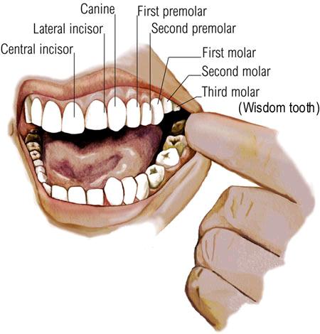
There are several types of teeth. Incisors are the squarish, sharp-edged teeth in the front of the mouth. There are four on the bottom and four on the top. On either side of the incisors are the sharp canines. The upper canines are sometimes called eyeteeth. Behind the canines are the premolars, or bicuspids. There are two sets, or four premolars, in each jaw.

The molars, situated behind the premolars, have points and grooves. There are 12 molars — three sets in each jaw called the first, second, and third molars. The third molars are the wisdom teeth, thought by some to have evolved thousands of years ago when human diets consisted of mostly raw foods that required extra chewing power. But because they can crowd out the other teeth, sometimes a dentist will need to remove them.

Human teeth are made up of four different types of tissue: pulp, dentin, enamel, and cementum. The pulp is the innermost portion of the tooth and consists of connective tissue, nerves, and blood vessels, which nourish the tooth. The pulp has two parts — the pulp chamber, which lies in the crown, and the root canal, which is in the root of the tooth. Blood vessels and nerves enter the root through a small hole in its tip and extend through the canal into the pulp chamber.

Dentin surrounds the pulp. A hard yellow substance consisting mostly of mineral salts and water, it makes up most of the tooth and is as hard as bone. It's the dentin that gives teeth their yellowish tint. Enamel, the hardest tissue in the body, covers the dentin and forms the outermost layer of the crown. It enables the tooth to withstand the pressure of chewing and protects it from harmful bacteria and changes in temperature from hot and cold foods. Both the dentin and pulp extend into the root. A bony layer of cementum covers the outside of the root, under the gum line, and holds the tooth in place within the jawbone. Cementum is also as hard as bone.

Normal Development of the Mouth and Teeth
Humans are diphyodont, meaning that they develop two sets of teeth. The first set of 20 deciduous teeth are also called the milk, primary, temporary, falling-off, or baby teeth. They begin to develop before birth and begin to fall out when a child is around 6 years old. They're replaced by a set of 32 permanent teeth, which are also called secondary or adult teeth.

Around the 8th week after conception, oval-shaped tooth buds consisting of cells form in the embryo. These buds begin to harden about the 16th week. Although teeth aren't visible at birth, both the primary and permanent teeth are forming below the gums. The crown, or the hard enamel-covered part that's visible in the mouth, develops first. When the crown is fully grown, the root begins to develop.

Between the ages of 6 months and 1 year, the deciduous teeth begin to push through the gums. This process is called eruption or teething. At this point, the crown is complete and the root is almost fully formed. By the time a child is 3 years old, he or she has a set of 20 deciduous teeth, 10 in the lower and 10 in the upper jaw. Each jaw has four incisors, two canines, and four molars. The molars' purpose is to grind food, and the incisors and canine teeth are used to bite into and tear food.

The primary teeth help the permanent teeth erupt in their normal positions; most of the permanent teeth form close to the roots of the primary teeth. When a primary tooth is preparing to fall out, its root begins to dissolve. This root has completely dissolved by the time the permanent tooth below it is ready to erupt.

Kids start to lose their primary teeth, or baby teeth, at about 6 years old. This begins a phase of permanent tooth development that lasts over the next 15 years, as the jaw steadily grows into its adult form. From ages 6 to 9, the incisors and first molars start to come in. Between ages 10 and 12, the first and second premolars, as well as the canines, erupt. From 12 to 13, the second molars come in. The wisdom teeth (third molars) erupt between the ages of 17 and 21.

Sometimes there isn't room in a person's mouth for all the permanent teeth. If this happens, the wisdom teeth may not come through at all. Overcrowding of the teeth is one of the reasons kids get braces.
What the Mouth and Teeth Do

The first step of digestion involves the mouth and teeth. Food enters the mouth and is immediately broken down into smaller pieces by our teeth. Each type of tooth serves a different function in the chewing process. Incisors cut foods when you bite into them. The sharper and longer canines tear food. The premolars, which are flatter than the canines, grind and mash food. Molars, with their points and grooves, are responsible for the most vigorous chewing. All the while, the tongue helps to push the food up against our teeth.

During chewing salivary glands in the walls and floor of the mouth secrete saliva, which moistens the food and helps break it down even more. Saliva makes it easier to chew and swallow foods (especially dry foods), and it contains enzymes that aid in the digestion of carbohydrates.

Once food has been converted into a soft, moist mass, it's pushed into the throat (or pharynx) at the back of the mouth and is swallowed. When we swallow, the soft palate closes off the nasal passages from the throat to prevent food from entering the nose.

Metabolism


Metabolism Basics

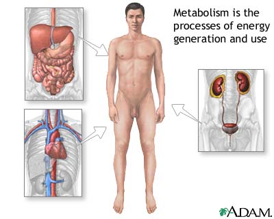
Specific proteins in the body control the chemical reactions of metabolism, and each chemical reaction is coordinated with other body functions. In fact, thousands of metabolic reactions happen at the same time — all regulated by the body — to keep our cells healthy and working.

Metabolism is a constant process that begins when we're conceived and ends when we die. It is a vital process for all life forms — not just humans. If metabolism stops, a living thing dies.

Here's an example of how the process of metabolism works in humans — and it begins with plants: First, a green plant takes in energy from sunlight. The plant uses this energy and the molecule cholorophyll (which gives plants their green color) to build sugars from water and carbon dioxide in a process known as photosynthesis.

When people and animals eat the plants (or, if they're carnivores, when they eat animals that have eaten the plants), they take in this energy (in the form of sugar), along with other vital cell-building chemicals. The body's next step is to break the sugar down so that the energy released can be distributed to, and used as fuel by, the body's cells.

After food is eaten, molecules in the digestive system called enzymes break proteins down into amino acids, fats into fatty acids, and carbohydrates into simple sugars (e.g., glucose). In addition to sugar, both amino acids and fatty acids can be used as energy sources by the body when needed. These compounds are absorbed into the blood, which transports them to the cells.

After they enter the cells, other enzymes act to speed up or regulate the chemical reactions involved with "metabolizing" these compounds. During these processes, the energy from these compounds can be released for use by the body or stored in body tissues, especially the liver, muscles, and body fat.

In this way, the process of metabolism is really a balancing act involving two kinds of activities that go on at the same time — the building up of body tissues and energy stores and the breaking down of body tissues and energy stores to generate more fuel for body functions.
  • Anabolism, or constructive metabolism, is all about building and storing: It supports the growth of new cells, the maintenance of body tissues, and the storage of energy for use in the future. During anabolism, small molecules are changed into larger, more complex molecules of carbohydrate, protein, and fat.
  • Catabolism, or destructive metabolism, is the process that produces the energy required for all activity in the cells. In this process, cells break down large molecules (mostly carbohydrates and fats) to release energy. This energy release provides fuel for anabolism, heats the body, and enables the muscles to contract and the body to move. As complex chemical units are broken down into more simple substances, the waste products released in the process of catabolism are removed from the body through the skin, kidneys, lungs, and intestines.

Several of the hormones of the endocrine system are involved in controlling the rate and direction of metabolism. Thyroxine, a hormone produced and released by the thyroid gland, plays a key role in determining how fast or slow the chemical reactions of metabolism proceed in a person's body.

Another gland, the pancreas secretes hormones that help determine whether the body's main metabolic activity at a particular time will be anabolic or catabolic. For example, after eating a meal, usually more anabolic activity occurs because eating increases the level of glucose — the body's most important fuel — in the blood. The pancreas senses this increased level of glucose and releases the hormone insulin, which signals cells to increase their anabolic activities.

Metabolism is a complicated chemical process, so it's not surprising that many people think of it in its simplest sense: as something that influences how easily our bodies gain or lose weight. That's where calories come in. A calorie is a unit that measures how much energy a particular food provides to the body. A chocolate bar has more calories than an apple, so it provides the body with more energy — and sometimes that can be too much of a good thing. Just as a car stores gas in the gas tank until it is needed to fuel the engine, the body stores calories — primarily as fat. If you overfill a car's gas tank, it spills over onto the pavement. Likewise, if a person eats too many calories, they "spill over" in the form of excess body fat.

The number of calories someone burns in a day is affected by how much that person exercises, the amount of fat and muscle in his or her body, and the person's basal metabolic rate (or BMR). BMR is a measure of the rate at which a person's body "burns" energy, in the form of calories, while at rest. The BMR can play a role in someone's tendency to gain weight. For example, a person with a low BMR (who therefore burns fewer calories while at rest or sleeping) will tend to gain more pounds of body fat over time, compared with a similar-sized person with an average BMR who eats the same amount of food and gets the same amount of exercise.

What factors influence BMR? To a certain extent, BMR is inherited. Sometimes health problems can affect BMR. But people can actually change their BMR in certain ways. For example, exercising more will not only cause a person to burn more calories directly from the extra activity itself, but becoming more physically fit will increase BMR as well. BMR is also influenced by body composition — people with more muscle and less fat generally have higher BMRs.

Lungs and Respiratory System


About the Lungs and Respiratory System
At the top of the respiratory system, the nostrils (also called nares) act as the air intake, bringing air into the nose, where it's warmed and humidified. Tiny hairs called cilia protect the nasal passageways and other parts of the respiratory tract, filtering out dust and other particles that enter the nose through the breathed air.

Air can also be taken in through the mouth. These two openings of the airway (the nasal cavity and the mouth) meet at the pharynx, or throat, at the back of the nose and mouth. The pharynx is part of the digestive system as well as the respiratory system because it carries both food and air. At the bottom of the pharynx, this pathway divides in two, one for food (the esophagus, which leads to the stomach) and the other for air. The epiglottis, a small flap of tissue, covers the air-only passage when you swallow, keeping food and liquid from going into the lungs.

The larynx, or voice box, is the uppermost part of the air-only pipe. This short tube contains a pair of vocal cords, which vibrate to make sounds. The trachea, or windpipe, extends downward from the base of the larynx. It lies partly in the neck and partly in the chest cavity. The walls of the trachea are strengthened by stiff rings of cartilage to keep it open. The trachea is also lined with cilia, which sweep fluids and foreign particles out of the airway so that they stay out of the lungs.

At its bottom end, the trachea divides into left and right air tubes called bronchi, which connect to the lungs. Within the lungs, the bronchi branch into smaller bronchi and even smaller tubes called bronchioles. Bronchioles end in tiny air sacs called alveoli, where the exchange of oxygen and carbon dioxide actually takes place. Each lung houses about 300 to 400 million alveoli. The lungs also contain elastic tissues that allow them to inflate and deflate without losing shape and are encased by a thin lining called the pleura. This network of alveoli, bronchioles, and bronchi is known as the bronchial tree.

The chest cavity, or thorax, is the airtight box that houses the bronchial tree, lungs, heart, and other structures. The top and sides of the thorax are formed by the ribs and attached muscles, and the bottom is formed by a large muscle called the diaphragm. The chest walls form a protective cage around the lungs and other contents of the chest cavity. Separating the chest from the abdomen, the diaphragm plays a lead role in breathing. It moves downward when we breathe in, enlarging the chest cavity and pulling air in through the nose or mouth. When we breathe out, the diaphragm moves upward, forcing the chest cavity to get smaller and pushing the gases in the lungs up and out of the nose and mouth.

What the Lungs and Respiratory System Do
The air we breathe is made up of several gases. Oxygen is the most important for keeping us alive because body cells need it for energy and growth. Without oxygen, the body's cells would die.

Carbon dioxide is the waste gas produced when carbon is combined with oxygen as part of the energy-making processes of the body. The lungs and respiratory system allow oxygen in the air to be taken into the body, while also enabling the body to get rid of carbon dioxide in the air breathed out.

Respiration is the set of events that results in the exchange of oxygen from the environment and carbon dioxide from the body's cells. The process of taking air into the lungs is inspiration, or inhalation, and the process of breathing it out is expiration, or exhalation.

Air is inhaled through the mouth or through the nose. Cilia lining the nose and other parts of the upper respiratory tract move back and forth, pushing foreign matter that comes in with air (like dust) either toward the nostrils to be expelled or toward the pharynx. The pharynx passes the foreign matter along to the stomach to eventually be eliminated by the body. As air is inhaled, the mucous membranes of the nose and mouth warm and humidify the air before it enters the lungs.

When you breathe in, the diaphragm moves downward toward the abdomen, and the rib muscles pull the ribs upward and outward. In this way, the volume of the chest cavity is increased. Air pressure in the chest cavity and lungs is reduced, and because gas flows from high pressure to low, air from the environment flows through the nose or mouth into the lungs. In exhalation, the diaphragm moves upward and the chest wall muscles relax, causing the chest cavity to contract. Air pressure in the lungs rises, so air flows from the lungs and up and out of respiratory system through the nose or mouth.

Every few seconds, with each inhalation, air fills a large portion of the millions of alveoli. In a process called diffusion, oxygen moves from the alveoli to the blood through the capillaries (tiny blood vessels) lining the alveolar walls. Once in the bloodstream, oxygen gets picked up by the hemoglobin in red blood cells. This oxygen-rich blood then flows back to the heart, which pumps it through the arteries to oxygen-hungry tissues throughout the body. In the tiny capillaries of the body tissues, oxygen is freed from the hemoglobin and moves into the cells. Carbon dioxide, which is produced during the process of diffusion, moves out of these cells into the capillaries, where most of it is dissolved in the plasma of the blood. Blood rich in carbon dioxide then returns to the heart via the veins. From the heart, this blood is pumped to the lungs, where carbon dioxide passes into the alveoli to be exhaled.

Kidneys and Urinary Tract


Our bodies produce several kinds of wastes, including sweat, carbon dioxide gas, feces (stool), and urine. These wastes exit the body in different ways. Sweat is released through pores in the skin. Water vapor and carbon dioxide are exhaled from the lungs. And undigested food materials are formed into feces in the intestines and excreted from the body as solid waste in bowel movements.

Urine, which is produced by the kidneys, contains the byproducts of metabolism — salts, toxins, and water — that end up in the blood. The kidneys and urinary tract (which includes the kidneys, ureters, bladder, and urethra) filter and eliminate these waste substances from our blood. Without the kidneys, waste products and toxins would soon build up in the blood to dangerous levels.

In addition to eliminating wastes, the kidneys and urinary tract also regulate many important body functions. For example, the kidneys monitor and maintain the body's balance of water, ensuring that our tissues receive enough water to function properly and be healthy.

When doctors take a urine sample, the results reveal how well the kidneys are working. For example, blood, protein, or white blood cells in the urine may indicate injury, inflammation, or infection of the kidneys, and glucose in the urine may be an indication of diabetes.

What the Kidneys and Urinary Tract Do

Although the two kidneys work together to perform many vital functions, people can live a normal, healthy life with just one kidney. In fact, some people are born with just one of these bean-shaped organs. If one kidney is removed, the remaining one will enlarge within a few months to take over the role of filtering blood on its own.

Every minute, more than 1 quart (about 1 liter) of blood goes to the kidneys. About one fifth of the blood pumped from the heart goes to the kidneys at any one time.

In addition to filtering blood, producing urine, and ensuring that body tissues receive enough water, the kidneys also regulate blood pressure and the level of vital salts in the blood. By regulating salt levels through production of an enzyme called renin (as well as other substances), the kidneys ensure that blood pressure is regulated.

The kidneys also secrete the hormone erythropoietin, which stimulates and controls red blood cell production (red blood cells carry oxygen throughout the body). In addition, the kidneys help regulate the acid-base balance (or the pH) of the blood and body fluids, which is necessary for the body to function normally.

How the Kidneys and Urinary Tract Work

The kidneys are located just under the ribcage in the back, one on each side. The right kidney is located below the liver, so it's a little lower than the left one. Each adult kidney is about the size of a fist. Each has an outer layer called the cortex, which contains the filtering units.

The center part of the kidney, the medulla has 10 to 15 fan-shaped structures called pyramids. These drain urine into cup-shaped tubes called calyxes. A layer of fat surrounds the kidneys to cushion and help hold them in place.

Here's how the kidneys filter blood : Blood travels to each kidney through the renal artery, which enters the kidney at the hilus, the indentation in the kidney that gives it its bean shape. As it enters the cortex, the artery branches to envelope the nephrons — 1 million tiny filtering units in each kidney that remove the harmful substances from the blood.

Each of the nephrons contain a filter called the glomerulus, which contains a network of tiny blood vessels known as capillaries. The fluid filtered from the blood by the glomerulus then travels down a tiny tube-like structure called a tubule, which adjusts the level of salts, water, and wastes that are excreted in the urine.

Filtered blood leaves the kidney through the renal vein and flows back to the heart.

The continuous blood supply entering and leaving the kidneys gives the kidneys their dark red color. While the blood is in the kidneys, water and some of the other blood components (such as acids, glucose, and other nutrients) are reabsorbed back into the bloodstream. Left behind is urine. Urine is a concentrated solution of waste material containing water, urea, a waste product that forms when proteins are broken down), salts, amino acids, byproducts of bile from the liver, ammonia, and any substances that cannot be reabsorbed into the blood. Urine also contains urochrome, a pigmented blood product that gives urine its yellowish color.

The renal pelvis, located near the hilus, collects the urine flowing from the calyxes. From the renal pelvis, urine is transported out of the kidneys through the ureters, tubes that carry the urine out of each kidney to be stored in the urinary bladder — a muscular collection sac in the lower abdomen.

The bladder expands as it fills and can hold about 2 cups (half a liter) of urine at any given time (an average adult produces about 6 cups, or 1½ liters, of urine per day). An adult needs to produce and excrete at least one third of this amount in order to adequately clear waste products from the body. Producing too much or not enough urine may indicate illness.

When the bladder is full, nerve endings in its wall send impulses to the brain. When a person is ready to urinate, the bladder walls contract and the sphincter (a ring-like muscle that guards the exit from the bladder to the urethra) relaxes. The urine is ejected from the bladder and out of the body through the urethra, another tube-like structure. The male urethra ends at the tip of the penis; the female urethra ends just above the vaginal opening.

Immune System


The immune system, which is made up of special cells, proteins, tissues, and organs, defends people against germs and microorganisms every day. In most cases, the immune system does a great job of keeping people healthy and preventing infections. But sometimes problems with the immune system can lead to illness and infection.

What the Immune System Does

The immune system is the body's defense against infectious organisms and other invaders. Through a series of steps called the immune response, the immune system attacks organisms and substances that invade our systems and cause disease. The immune system is made up of a network of cells, tissues, and organs that work together to protect the body.

The cells that are part of this defense system are white blood cells, or leukocytes. They come in two basic types (more on these below), which combine to seek out and destroy the organisms or substances that cause disease.

Leukocytes are produced or stored in many locations throughout the body, including the thymus, spleen, and bone marrow. For this reason, they are called the lymphoid organs. There are also clumps of lymphoid tissue throughout the body, primarily in the form of lymph nodes, that house the leukocytes.

The leukocytes circulate through the body between the organs and nodes by means of the lymphatic vessels. Leukocytes can also circulate through the blood vessels. In this way, the immune system works in a coordinated manner to monitor the body for germs or substances that might cause problems.

The two basic types of leukocytes are:
  • phagocytes, cells that chew up invading organisms
  • lymphocytes, cells that allow the body to remember and recognize previous invaders and help the body destroy them

A number of different cells are considered phagocytes. The most common type is the neutrophil, which primarily fights bacteria. If doctors are worried about a bacterial infection, they might order a blood test to see if a patient has an increased number of neutrophils triggered by the infection. Other types of phagocytes have their own jobs to make sure that the body responds appropriately to a specific type of invader.

There are two kinds of lymphocytes: the B lymphocytes and the T lymphocytes. Lymphocytes start out in the bone marrow and either stay there and mature into B cells, or they leave for the thymus gland, where they mature into T cells. B lymphocytes and T lymphocytes have separate jobs to do: B lymphocytes are like the body's military intelligence system, seeking out their targets and sending defenses to lock onto them. T cells are like the soldiers, destroying the invaders that the intelligence system has identified. Here's how it works.

Antigens are foreign substances that invade the body. When an antigen is detected, several types of cells work together to recognize and respond to it. These cells trigger the B lymphocytes to produce antibodies, specialized proteins that lock onto specific antigens. Antibodies and antigens fit together like a key and a lock.

Once the B lymphocytes have produced antibodies, these antibodies continue to exist in a person's body, so that if the same antigen is presented to the immune system again, the antibodies are already there to do their job. That's why if someone gets sick with a certain disease, like chickenpox, that person typically doesn't get sick from it again. This is also why we use immunizations to prevent getting certain diseases. The immunization introduces the body to the antigen in a way that doesn't make a person sick, but it does allow the body to produce antibodies that will then protect that person from future attack by the germ or substance that produces that particular disease.

Although antibodies can recognize an antigen and lock onto it, they are not capable of destroying it without help. That is the job of the T cells. The T cells are part of the system that destroys antigens that have been tagged by antibodies or cells that have been infected or somehow changed. (There are actually T cells that are called "killer cells.") T cells are also involved in helping signal other cells (like phagocytes) to do their jobs.

Antibodies can also neutralize toxins (poisonous or damaging substances) produced by different organisms. Lastly, antibodies can activate a group of proteins called complement that are also part of the immune system. Complement assists in killing bacteria, viruses, or infected cells.

All of these specialized cells and parts of the immune system offer the body protection against disease. This protection is called immunity.

Humans have three types of immunity — innate, adaptive, and passive:

Innate Immunity

Everyone is born with innate (or natural) immunity, a type of general protection that humans have. Many of the germs that affect other species don't harm us. For example, the viruses that cause leukemia in cats or distemper in dogs don't affect humans. Innate immunity works both ways because some viruses that make humans ill — such as the virus that causes HIV/AIDS — don't make cats or dogs sick either.

Innate immunity also includes the external barriers of the body, like the skin and mucous membranes (like those that line the nose, throat, and gastrointestinal tract), which are our first line of defense in preventing diseases from entering the body. If this outer defensive wall is broken (like if you get a cut), the skin attempts to heal the break quickly and special immune cells on the skin attack invading germs.

Adaptive Immunity

We also have a second kind of protection called adaptive (or active) immunity. This type of immunity develops throughout our lives. Adaptive immunity involves the lymphocytes (as in the process described above) and develops as children and adults are exposed to diseases or immunized against diseases through vaccination.

Passive Immunity

Passive immunity is "borrowed" from another source and it lasts for a short time. For example, antibodies in a mother's breast milk provide an infant with temporary immunity to diseases that the mother has been exposed to. This can help protect the infant against infection during the early years of childhood.

Everyone's immune system is different. Some people never seem to get infections, whereas others seem to be sick all the time. As people get older, they usually become immune to more germs as the immune system comes into contact with more and more of them. That's why adults and teens tend to get fewer colds than kids — their bodies have learned to recognize and immediately attack many of the viruses that cause colds.

Heart and Circulatory System


About the Heart and Circulatory System

The circulatory system is composed of the heart and blood vessels, including arteries, veins, and capillaries. Our bodies actually have two circulatory systems: The pulmonary circulation is a short loop from the heart to the lungs and back again, and the systemic circulation (the system we usually think of as our circulatory system) sends blood from the heart to all the other parts of our bodies and back again.

The heart is the key organ in the circulatory system. As a hollow, muscular pump, its main function is to propel blood throughout the body. It usually beats from 60 to 100 times per minute, but can go much faster when necessary. It beats about 100,000 times a day, more than 30 million times per year, and about 2.5 billion times in a 70-year lifetime.

The heart gets messages from the body that tell it when to pump more or less blood depending on an individual's needs. When we're sleeping, it pumps just enough to provide for the lower amounts of oxygen needed by our bodies at rest. When we're exercising or frightened, the heart pumps faster to increase the delivery of oxygen.

The heart has four chambers that are enclosed by thick, muscular walls. It lies between the lungs and just to the left of the middle of the chest cavity. The bottom part of the heart is divided into two chambers called the right and left ventricles, which pump blood out of the heart. A wall called the interventricular septum divides the ventricles.

The upper part of the heart is made up of the other two chambers of the heart, the right and left atria. The right and left atria receive the blood entering the heart. A wall called the interatrial septum divides the right and left atria, which are separated from the ventricles by the atrioventricular valves. The tricuspid valve separates the right atrium from the right ventricle, and the mitral valve separates the left atrium and the left ventricle.

Two other cardiac valves separate the ventricles and the large blood vessels that carry blood leaving the heart. These are the pulmonic valve, which separates the right ventricle from the pulmonary artery leading to the lungs, and the aortic valve, which separates the left ventricle from the aorta, the body's largest blood vessel.

Arteries carry blood away from the heart. They are the thickest blood vessels, with muscular walls that contract to keep the blood moving away from the heart and through the body. In the systemic circulation, oxygen-rich blood is pumped from the heart into the aorta. This huge artery curves up and back from the left ventricle, then heads down in front of the spinal column into the abdomen. Two coronary arteries branch off at the beginning of the aorta and divide into a network of smaller arteries that provide oxygen and nourishment to the muscles of the heart.

Unlike the aorta, the body's other main artery, the pulmonary artery, carries oxygen-poor blood. From the right ventricle, the pulmonary artery divides into right and left branches, on the way to the lungs where blood picks up oxygen.

Arterial walls have three layers:
  1. The endothelium is on the inside and provides a smooth lining for blood to flow over as it moves through the artery.
  2. The media is the middle part of the artery, made up of a layer of muscle and elastic tissue.
  3. The adventitia is the tough covering that protects the outside of the artery.

As they get farther from the heart, the arteries branch out into arterioles, which are smaller and less elastic.

Veins carry blood back to the heart. They're not as muscular as arteries, but they contain valves that prevent blood from flowing backward. Veins have the same three layers that arteries do, but are thinner and less flexible. The two largest veins are the superior and inferior vena cavae. The terms superior and inferior don't mean that one vein is better than the other, but that they're located above and below the heart.

A network of tiny capillaries connects the arteries and veins. Though tiny, the capillaries are one of the most important parts of the circulatory system because it's through them that nutrients and oxygen are delivered to the cells. In addition, waste products such as carbon dioxide are also removed by the capillaries.

What the Heart and Circulatory System Do

The circulatory system works closely with other systems in our bodies. It supplies oxygen and nutrients to our bodies by working with the respiratory system. At the same time, the circulatory system helps carry waste and carbon dioxide out of the body.

Hormones — produced by the endocrine system — are also transported through the blood in the circulatory system. As the body's chemical messengers, hormones transfer information and instructions from one set of cells to another. For example, one of the hormones produced by the heart helps control the kidneys' release of salt from the body.

One complete heartbeat makes up a cardiac cycle, which consists of two phases:
  1. In the first phase, the ventricles contract (this is called systole), sending blood into the pulmonary and systemic circulation. To prevent the flow of blood backwards into the atria during systole, the atrioventricular valves close, creating the first sound (the lub). When the ventricles finish contracting, the aortic and pulmonary valves close to prevent blood from flowing back into the ventricles. This is what creates the second sound (the dub).
  2. Then the ventricles relax (this is called diastole) and fill with blood from the atria, which makes up the second phase of the cardiac cycle.

A unique electrical conduction system in the heart causes it to beat in its regular rhythm. The sinoatrial or SA node, a small area of tissue in the wall of the right atrium, sends out an electrical signal to start the contracting of the heart muscle. This node is called the pacemaker of the heart because it sets the rate of the heartbeat and causes the rest of the heart to contract in its rhythm.

These electrical impulses cause the atria to contract first, and then travel down to the atrioventricular or AV node, which acts as a kind of relay station. From here the electrical signal travels through the right and left ventricles, causing them to contract and forcing blood out into the major arteries.

In the systemic circulation, blood travels out of the left ventricle, to the aorta, to every organ and tissue in the body, and then back to the right atrium. The arteries, capillaries, and veins of the systemic circulatory system are the channels through which this long journey takes place.

Once in the arteries, blood flows to smaller arterioles and then to capillaries. While in the capillaries, the bloodstream delivers oxygen and nutrients to the body's cells and picks up waste materials. Blood then goes back through the capillaries into venules, and then to larger veins until it reaches the vena cavae.

Blood from the head and arms returns to the heart through the superior vena cava, and blood from the lower parts of the body returns through the inferior vena cava. Both vena cavae deliver this oxygen-depleted blood into the right atrium. From here the blood exits to fill the right ventricle, ready to be pumped into the pulmonary circulation for more oxygen.

In the pulmonary circulation, blood low in oxygen but high in carbon dioxide is pumped out the right ventricle into the pulmonary artery, which branches off in two directions. The right branch goes to the right lung, and vice versa.

In the lungs, the branches divide further into capillaries. Blood flows more slowly through these tiny vessels, allowing time for gases to be exchanged between the capillary walls and the millions of alveoli, the tiny air sacs in the lungs.

During the process called oxygenation, oxygen is taken up by the bloodstream. Oxygen locks onto a molecule called hemoglobin in the red blood cells. The newly oxygenated blood leaves the lungs through the pulmonary veins and heads back to the heart. It enters the heart in the left atrium, then fills the left ventricle so it can be pumped into the systemic circulation.

Eyes


We depend on sight more than any other of our senses to maneuver through the space around us. In a single glance, lasting a fraction of a second, our eyes work with our brains to tell us the size, shape, color, and texture of an object. They let us know how close it is, whether it's standing still or coming toward us, and how quickly it's moving. Every day, our eyes give us messages that help us understand the world around us.

Although the eyes are small compared with most of the body's other organs, their structure is incredibly complex. The eyes work together to perceive depth, enabling us to judge distance and the size of objects to help us move around them. Not only do the two eyes work together, they also work with the brain, muscles, and nerves to produce complicated visual images and messages. And our eyes constantly adapt to the changing environment — for example, they are able to adjust so that we can easily move around in a nearly dark room or bright sunlight.

To understand more about how the eyes work, it's important to know about the structures that make up the eye and about conditions and diseases that can interfere with vision.

Eyes and What Do They Do

Only part of the eye is visible in a person's face. The whole eye — the eyeball — is about the size and shape of a ping-pong ball.

The eye — both the parts that are visible and those that aren't — is extremely delicate. The body has several ways of protecting this vulnerable organ. The eyeball sits in the eye socket (also called the orbit) in a person's skull, where it is surrounded by bone. The visible part of the eye is protected by the eyelids and the eyelashes, which keep dirt, dust, and even harmful bright light out of the eye.

Our eyes are also protected by tears, which moisten the eyes and clean out dirt, dust, and other irritants that get past the defenses of our eyelashes and eyelids. Tears also help protect the eyes against infection.

Every time we blink, our eyelids spread a layer of mucus, oil, and tears over the cornea, which covers the eye. The lacrimal glands in the upper outer corner of each eye socket produce tears.

After they've done their job moistening the eyes, the tears flow into canals in the eyelids, which drain into the lacrimal sac, a pouch in the lower inner corner of each eye socket. Tears then exit through a passage which leads to the nose.

To see, the eye has to move. Six muscles, called extraocular muscles, surround the eyeball in the skull. These muscles act like the strings on a puppet, moving the eye in different directions. The muscles of each eye normally move together at the same time, allowing the two eyes to remain aligned.

The wall of a person's eyeball is made up of three layers, rather like the layers on an onion:
  • The sclera is the outermost protective layer. This tough, fibrous tissue surrounds the eyeball and attaches to the cornea, which is the clear front surface of the eye. What we see as the white of the eye is the sclera. Over the sclera lies the conjunctiva, a clear mucous membrane that protects the eye from becoming dry.
  • The choroid is the middle layer that contains blood vessels that deliver oxygen and nutrients to the retina.
  • The retina, the innermost of the three layers, lines the inside of the eyeball. The retina is a soft, light-sensitive layer of nervous system tissue. The optic nerve carries signals from the retina to the brain, which interprets them as visual images.

The space in the center of the eyeball is filled with a clear jelly-like material called the vitreous humor. This material allows light to pass through to the retina. It also helps the eye keep its round shape.

Vision is the process by which images captured by the eye are interpreted by the brain, and the visible part of the eye is where the process of sight begins. On the front surface of the eye is the see-through, circle-shaped cornea. You can't see a person's cornea the way you can see the colored part of the eye behind it — the cornea is like a clear window that focuses light into the eye. Behind the cornea is a watery fluid called the aqueous humor. The cornea and aqueous humor form an outer lens that refracts (bends) light on its way into the eye. This is where most of the eye's focusing work is done.

The colored circular membrane in the eye just behind the cornea is called the iris. The iris controls the amount of light entering the eye through the pupil, which is the opening in the center of the iris that looks like a tiny black circle. Like a camera, which controls the amount of light coming in to prevent both overexposure and underexposure, the iris becomes wider and narrower, changing the size of the pupil to control the amount of light entering the eye. The pupil gets bigger when more light is needed to see better and smaller when there's plenty of light.

The eye's lens sits just behind the iris. Just like a camera lens, the eye's lens focuses light to form sharp, clear images. Light that has been focused through the cornea and aqueous humor hits the lens, which then focuses it further, sending the light rays through the vitreous humor and onto the retina.

To focus on objects clearly at varying distances, the eye's lens needs to change shape. The ciliary body contains the muscular structure in the eye that changes the shape of the eye's lens. In people who have normal vision, the ciliary body flattens the lens enough to bring objects into focus at a distance of 20 feet or more. To see closer objects, this muscle contracts to thicken the lens. Young children can see objects at very close range; many people over 45 have to hold objects farther and farther away to see them clearly. This is because the lens becomes less elastic as we age.

The retina (the soft, light-sensitive layer of tissue that lines the back of the eyeball wall) is made up of millions of light receptors. These are called rods and cones. Rods are much more sensitive to light than cones. Each eye has about 125 million rods that help us see in dim light and detect shades of gray, but they cannot distinguish colors. In comparison, the 6 million cones in each eye allow us to see in bright light, and they also sense color and detail.

The macula is a small, specialized area on the retina. The macula helps our eyes see fine details when we look directly at an object. It contains mainly cones and few rods.

When focused light is projected onto the retina, it stimulates the rods and cones. The retina then sends nerve signals are sent through the back of the eye to the optic nerve. The optic nerve carries these signals to the brain, which interprets them as visual images. The portion of the brain that processes visual input and interprets the messages that the eye sends is called the visual cortex.

As in a camera, the eye's lens transmits light patterns upside down. The brain learns that the impulses received from the upper part of the retina are really from the lower part of the object we're seeing and vice versa.

Most people use both eyes to see an object. This is called binocular vision. Through binocular vision, images are formed on the retina of each eye. These images are slightly different, because the object is being viewed from slightly different angles. Nerve signals representing each image are sent to the brain, where they are interpreted as two views of the same object. Some of the nerve fibers from each eye cross, so each side of the brain receives messages from both eyes. Through experience, the brain learns to judge the distance of an object by the degree of difference in the images it receives from the two eyes. This ability to sense distance is called depth perception.

Vision is a fine-tuned process. All the parts of the eye — and the brain — need to work together so a person can see correctly. Because the eye's structure is so complex, though, a lot of things can go wrong.

Endocrine System


The foundations of the endocrine system are the hormones and glands. As the body's chemical messengers, hormones transfer information and instructions from one set of cells to another. Although many different hormones circulate throughout the bloodstream, each one affects only the cells that are genetically programmed to receive and respond to its message. Hormone levels can be influenced by factors such as stress, infection, and changes in the balance of fluid and minerals in blood.

A gland is a group of cells that produces and secretes, or gives off, chemicals. A gland selects and removes materials from the blood, processes them, and secretes the finished chemical product for use somewhere in the body. Some types of glands release their secretions in specific areas. For instance, exocrine glands, such as the sweat and salivary glands, release secretions in the skin or inside of the mouth. Endocrine glands, on the other hand, release more than 20 major hormones directly into the bloodstream where they can be transported to cells in other parts of the body.

The major glands that make up the human endocrine system are the hypothalamus, pituitary, thyroid, parathyroids, adrenals, pineal body, and the reproductive glands, which include the ovaries and testes. The pancreas is also part of this hormone-secreting system, even though it is also associated with the digestive system because it also produces and secretes digestive enzymes.

Although the endocrine glands are the body's main hormone producers, some non-endocrine organs — such as the brain, heart, lungs, kidneys, liver, thymus, skin, and placenta — also produce and release hormones.

The hypothalamus, a collection of specialized cells that is located in the lower central part of the brain, is the primary link between the endocrine and nervous systems. Nerve cells in the hypothalamus control the pituitary gland by producing chemicals that either stimulate or suppress hormone secretions from the pituitary.

Although it is no bigger than a pea, the pituitary gland, located at the base of the brain just beneath the hypothalamus, is considered the most important part of the endocrine system. It's often called the "master gland" because it makes hormones that control several other endocrine glands. The production and secretion of pituitary hormones can be influenced by factors such as emotions and seasonal changes. To accomplish this, the hypothalamus relays information sensed by the brain (such as environmental temperature, light exposure patterns, and feelings) to the pituitary.

The tiny pituitary is divided into two parts: the anterior lobe and the posterior lobe. The anterior lobe regulates the activity of the thyroid, adrenals, and reproductive glands. Among the hormones it produces are:

  • growth hormone, which stimulates the growth of bone and other body tissues and plays a role in the body's handling of nutrients and minerals
  • prolactin, which activates milk production in women who are breastfeeding
  • thyrotropin, which stimulates the thyroid gland to produce thyroid hormones
  • corticotropin, which stimulates the adrenal gland to produce certain hormones

  • The pituitary also secretes endorphins, chemicals that act on the nervous system to reduce sensitivity to pain. In addition, the pituitary secretes hormones that signal the ovaries and testes to make sex hormones. The pituitary gland also controls ovulation and the menstrual cycle in women.

    The posterior lobe of the pituitary releases antidiuretic hormone, which helps control body water balance through its effect on the kidneys and urine output; and oxytocin, which triggers the contractions of the uterus that occur during labor.

    The thyroid, located in the front part of the lower neck, is shaped like a bowtie or butterfly and produces the thyroid hormones thyroxine and triiodothyronine. These hormones control the rate at which cells burn fuels from food to produce energy. As the level of thyroid hormones increases in the bloodstream, so does the speed at which chemical reactions occur in the body.

    Thyroid hormones also play a key role in bone growth and the development of the brain and nervous system in children. The production and release of thyroid hormones is controlled by thyrotropin, which is secreted by the pituitary gland.

    Attached to the thyroid are four tiny glands that function together called the parathyroids. They release parathyroid hormone, which regulates the level of calcium in the blood with the help of calcitonin, which is produced in the thyroid.

    The body has two triangular adrenal glands, one on top of each kidney. The adrenal glands have two parts, each of which produces a set of hormones and has a different function. The outer part, the adrenal cortex, produces hormones called corticosteroids that influence or regulate salt and water balance in the body, the body's response to stress, metabolism, the immune system, and sexual development and function.

    The inner part, the adrenal medulla, produces catecholamines, such as epinephrine. Also called adrenaline, epinephrine increases blood pressure and heart rate when the body experiences stress. (Epinephrine injections are often used to counteract a severe allergic reaction.)

    The pineal body, also called the pineal gland, is located in the middle of the brain. It secretes melatonin, a hormone that may help regulate the wake-sleep cycle.

    The gonads are the main source of sex hormones. In males, they are located in the scrotum. Male gonads, or testes, secrete hormones called androgens, the most important of which is testosterone. These hormones regulate body changes associated with sexual development, including enlargement of the penis, the growth spurt that occurs during puberty, and the appearance of other male secondary sex characteristics such as deepening of the voice, growth of facial and pubic hair, and the increase in muscle growth and strength. Working with hormones from the pituitary gland, testosterone also supports the production of sperm by the testes.

    The female gonads, the ovaries, are located in the pelvis. They produce eggs and secrete the female hormones estrogen and progesterone. Estrogen is involved in the development of female sexual features such as breast growth, the accumulation of body fat around the hips and thighs, and the growth spurt that occurs during puberty. Both estrogen and progesterone are also involved in pregnancy and the regulation of the menstrual cycle.

    The pancreas produces (in addition to others) two important hormones, insulin and glucagon. They work together to maintain a steady level of glucose, or sugar, in the blood and to keep the body supplied with fuel to produce and maintain stores of energy.

    What the Endocrine System Does

    Once a hormone is secreted, it travels from the endocrine gland through the bloodstream to target cells designed to receive its message. Along the way to the target cells, special proteins bind to some of the hormones. The special proteins act as carriers that control the amount of hormone that is available to interact with and affect the target cells.

    Also, the target cells have receptors that latch onto only specific hormones, and each hormone has its own receptor, so that each hormone will communicate only with specific target cells that possess receptors for that hormone. When the hormone reaches its target cell, it locks onto the cell's specific receptors and these hormone-receptor combinations transmit chemical instructions to the inner workings of the cell.

    When hormone levels reach a certain normal or necessary amount, further secretion is controlled by important body mechanisms to maintain that level of hormone in the blood. This regulation of hormone secretion may involve the hormone itself or another substance in the blood related to the hormone.

    For example, if the thyroid gland has secreted adequate amounts of thyroid hormones into the blood, the pituitary gland senses the normal levels of thyroid hormone in the bloodstream and adjusts its release of thyrotropin, the pituitary hormone that stimulates the thyroid gland to produce thyroid hormones.

    Another example is parathyroid hormone, which increases the level of calcium in the blood. When the blood calcium level rises, the parathyroid glands sense the change and decrease their secretion of parathyroid hormone. This turnoff process is called a negative feedback system.

    Digestive System


    What's the first step in the digestive process? Believe it or not, it happens before you even taste your food. Just by smelling that homemade apple pie or thinking about how delicious that ripe tomato is going to be, you start salivating — and the digestive process begins, preparing for that first scrumptious bite.

    Food is our fuel, and its nutrients give our bodies' cells the energy and substances they need to operate. But before food can do that, it must be digested into small pieces the body can absorb and use.

    About the Digestive System

    Almost all animals have a tube-type digestive system in which food enters the mouth, passes through a long tube, and exits as feces (poop) through the anus. The smooth muscle in the walls of the tube-shaped digestive organs rhythmically and efficiently moves the food through the system, where it is broken down into tiny absorbable atoms and molecules.

    During the process of absorption, nutrients that come from the food (including carbohydrates, proteins, fats, vitamins, and minerals) pass through channels in the intestinal wall and into the bloodstream. The blood works to distribute these nutrients to the rest of the body. The waste parts of food that the body can't use are passed out of the body as feces.

    Every morsel of food we eat has to be broken down into nutrients that can be absorbed by the body, which is why it takes hours to fully digest food. In humans, protein must be broken down into amino acids, starches into simple sugars, and fats into fatty acids and glycerol. The water in our food and drink is also absorbed into the bloodstream to provide the body with the fluid it needs.

    How Digestion Works

    The digestive system is made up of the alimentary canal (also called the digestive tract) and the other abdominal organs that play a part in digestion, such as the liver and pancreas. The alimentary canal is the long tube of organs — including the esophagus, stomach, and intestines — that runs from the mouth to the anus. An adult's digestive tract is about 30 feet (about 9 meters) long.

    Digestion begins in the mouth, well before food reaches the stomach. When we see, smell, taste, or even imagine a tasty meal, our salivary glands, which are located under the tongue and near the lower jaw, begin producing saliva. This flow of saliva is set in motion by a brain reflex that's triggered when we sense food or think about eating. In response to this sensory stimulation, the brain sends impulses through the nerves that control the salivary glands, telling them to prepare for a meal.

    As the teeth tear and chop the food, saliva moistens it for easy swallowing. A digestive enzyme called amylase, which is found in saliva, starts to break down some of the carbohydrates (starches and sugars) in the food even before it leaves the mouth.

    Swallowing, which is accomplished by muscle movements in the tongue and mouth, moves the food into the throat, or pharynx. The pharynx, a passageway for food and air, is about 5 inches (12.7 centimeters) long. A flexible flap of tissue called the epiglottis reflexively closes over the windpipe when we swallow to prevent choking.

    From the throat, food travels down a muscular tube in the chest called the esophagus. Waves of muscle contractions called peristalsis force food down through the esophagus to the stomach. A person normally isn't aware of the movements of the esophagus, stomach, and intestine that take place as food passes through the digestive tract.

    At the end of the esophagus, a muscular ring or valve called a sphincter allows food to enter the stomach and then squeezes shut to keep food or fluid from flowing back up into the esophagus. The stomach muscles churn and mix the food with acids and enzymes, breaking it into much smaller, digestible pieces. An acidic environment is needed for the digestion that takes place in the stomach. Glands in the stomach lining produce about 3 quarts (2.8 liters) of these digestive juices each day.

    Most substances in the food we eat need further digestion and must travel into the intestine before being absorbed. When it's empty, an adult's stomach has a volume of one fifth of a cup (1.6 fluid ounces), but it can expand to hold more than 8 cups (64 fluid ounces) of food after a large meal.

    By the time food is ready to leave the stomach, it has been processed into a thick liquid called chyme. A walnut-sized muscular valve at the outlet of the stomach called the pylorus keeps chyme in the stomach until it reaches the right consistency to pass into the small intestine. Chyme is then squirted down into the small intestine, where digestion of food continues so the body can absorb the nutrients into the bloodstream.

    The small intestine is made up of three parts:
    • the duodenum, the C-shaped first part
    • the jejunum, the coiled midsection
    • the ileum, the final section that leads into the large intestine

    The inner wall of the small intestine is covered with millions of microscopic, finger-like projections called villi. The villi are the vehicles through which nutrients can be absorbed into the body.

    The liver (located under the rib cage in the right upper part of the abdomen), the gallbladder (hidden just below the liver), and the pancreas (beneath the stomach) are not part of the alimentary canal, but these organs are essential to digestion.

    The liver produces bile, which helps the body absorb fat. Bile is stored in the gallbladder until it is needed. The pancreas produces enzymes that help digest proteins, fats, and carbohydrates. It also makes a substance that neutralizes stomach acid. These enzymes and bile travel through special channels (called ducts) directly into the small intestine, where they help to break down food. The liver also plays a major role in the handling and processing of nutrients, which are carried to the liver in the blood from the small intestine.

    From the small intestine, undigested food (and some water) travels to the large intestine through a muscular ring or valve that prevents food from returning to the small intestine. By the time food reaches the large intestine, the work of absorbing nutrients is nearly finished. The large intestine's main function is to remove water from the undigested matter and form solid waste that can be excreted. The large intestine is made up of these three parts:
    • The cecum is a pouch at the beginning of the large intestine that joins the small intestine to the large intestine. This transition area expands in diameter, allowing food to travel from the small intestine to the large. The appendix, a small, hollow, finger-like pouch, hangs at the end of the cecum. Doctors believe the appendix is left over from a previous time in human evolution. It no longer appears to be useful to the digestive process.
    • The colon extends from the cecum up the right side of the abdomen, across the upper abdomen, and then down the left side of the abdomen, finally connecting to the rectum. The colon has three parts: the ascending colon; the transverse colon, which absorb fluids and salts; and the descending colon, which holds the resulting waste. Bacteria in the colon help to digest the remaining food products.
    • The rectum is where feces are stored until they leave the digestive system through the anus as a bowel movement.

    Brain and Nervous System


    Anatomy of the Nervous System

    If you think of the brain as a central computer that controls all bodily functions, then the nervous system is like a network that relays messages back and forth from the brain to different parts of the body. It does this via the spinal cord, which runs from the brain down through the back and contains threadlike nerves that branch out to every organ and body part.

    When a message comes into the brain from anywhere in the body, the brain tells the body how to react. For example, if you accidentally touch a hot stove, the nerves in your skin shoot a message of pain to your brain. The brain then sends a message back telling the muscles in your hand to pull away. Luckily, this neurological relay race takes a lot less time than it just took to read about it.

    Considering everything it does, the human brain is incredibly compact, weighing just 3 pounds. Its many folds and grooves, though, provide it with the additional surface area necessary for storing all of the body's important information.

    The spinal cord, on the other hand, is a long bundle of nerve tissue about 18 inches long and ¾ inch thick. It extends from the lower part of the brain down through spine. Along the way, various nerves branch out to the entire body. These are called the peripheral nervous system.

    Both the brain and the spinal cord are protected by bone: the brain by the bones of the skull, and the spinal cord by a set of ring-shaped bones called vertebrae. They're both cushioned by layers of membranes called meninges as well as a special fluid called cerebrospinal fluid. This fluid helps protect the nerve tissue, keep it healthy, and remove waste products.

    The brain is made up of three main sections: the forebrain, the midbrain, and the hindbrain.

    The Forebrain
    The forebrain is the largest and most complex part of the brain. It consists of the cerebrum — the area with all the folds and grooves typically seen in pictures of the brain — as well as some other structures beneath it.

    The cerebrum contains the information that essentially makes us who we are: our intelligence, memory, personality, emotion, speech, and ability to feel and move. Specific areas of the cerebrum are in charge of processing these different types of information. These are called lobes, and there are four of them: the frontal, parietal, temporal, and occipital.

    The cerebrum has right and left halves, called hemispheres, which are connected in the middle by a band of nerve fibers (the corpus collosum) that enables the two sides to communicate. Though these halves may look like mirror images of each other, many scientists believe they have different functions. The left side is considered the logical, analytical, objective side. The right side is thought to be more intuitive, creative, and subjective. So when you're balancing the checkbook, you're using the left side; when you're listening to music, you're using the right side. It's believed that some people are more "right-brained" or "left-brained" while others are more "whole-brained," meaning they use both halves of their brain to the same degree.

    The outer layer of the cerebrum is called the cortex (also known as "gray matter"). Information collected by the five senses comes into the brain from the spinal cord to the cortex. This information is then directed to other parts of the nervous system for further processing. For example, when you touch the hot stove, not only does a message go out to move your hand but one also goes to another part of the brain to help you remember not to do that again.

    In the inner part of the forebrain sits the thalamus, hypothalamus, and pituitary gland. The thalamus carries messages from the sensory organs like the eyes, ears, nose, and fingers to the cortex. The hypothalamus controls the pulse, thirst, appetite, sleep patterns, and other processes in our bodies that happen automatically. It also controls the pituitary gland, which makes the hormones that control our growth, metabolism, digestion, sexual maturity, and response to stress.

    The Midbrain
    The midbrain, located underneath the middle of the forebrain, acts as a master coordinator for all the messages going in and out of the brain to the spinal cord.

    The Hindbrain
    The hindbrain sits underneath the back end of the cerebrum, and it consists of the cerebellum, pons, and medulla. The cerebellum — also called the "little brain" because it looks like a small version of the cerebrum — is responsible for balance, movement, and coordination.

    The pons and the medulla, along with the midbrain, are often called the brainstem. The brainstem takes in, sends out, and coordinates all of the brain's messages. It is also controls many of the body's automatic functions, like breathing, heart rate, blood pressure, swallowing, digestion, and blinking.

    How the Nervous System Works

    The basic functioning of the nervous system depends a lot on tiny cells called neurons. The brain has billions of them, and they have many specialized jobs. For example, sensory neurons take information from the eyes, ears, nose, tongue, and skin to the brain. Motor neurons carry messages away from the brain and back to the rest of the body.

    All neurons, however, relay information to each other through a complex electrochemical process, making connections that affect the way we think, learn, move, and behave.

    Intelligence, learning, and memory. At birth, the nervous system contains all the neurons you will ever have, but many of them are not connected to each other. As you grow and learn, messages travel from one neuron to another over and over, creating connections, or pathways, in the brain. It's why driving seemed to take so much concentration when you first learned but now is second nature: The pathway became established.

    In young children, the brain is highly adaptable; in fact, when one part of a young child's brain is injured, another part can often learn to take over some of the lost function. But as we age, the brain has to work harder to make new neural pathways, making it more difficult to master new tasks or change established behavior patterns. That's why many scientists believe it's important to keep challenging your brain to learn new things and make new connections— it helps keeps the brain active over the course of a lifetime.

    Memory is another complex function of the brain. The things we've done, learned, and seen are first processed in the cortex, and then, if we sense that this information is important enough to remember permanently, it's passed inward to other regions of the brain (such as the hippocampus and amygdala) for long-term storage and retrieval. As these messages travel through the brain, they too create pathways that serve as the basis of our memory.

    Movement. Different parts of the cerebrum are responsible for moving different body parts. The left side of the brain controls the movements of the right side of the body, and the right side of the brain controls the movements of the left side of the body. When you press the accelerator with your right foot, for example, it's the left side of your brain that sends the message allowing you to do it.

    Basic body functions. A part of the peripheral nervous system called the autonomic nervous system is responsible for controlling many of the body processes we almost never need to think about, like breathing, digestion, sweating, and shivering. The autonomic nervous system has two parts: the sympathetic and the parasympathetic nervous systems.

    The sympathetic nervous system prepares the body for sudden stress, like if you see a robbery taking place. When something frightening happens, the sympathetic nervous system makes the heart beat faster so that it sends blood more quickly to the different body parts that might need it. It also causes the adrenal glands at the top of the kidneys to release adrenaline, a hormone that helps give extra power to the muscles for a quick getaway. This process is known as the body's "fight or flight" response.

    The parasympathetic nervous system does the exact opposite: It prepares the body for rest. It also helps the digestive tract move along so our bodies can efficiently take in nutrients from the food we eat.

    The senses. Your spouse may be a sight for sore eyes at the end of a long day — but without the brain, you wouldn't even recognize him or her. Pepperoni pizza sure is delicious — but without the brain, your taste buds wouldn't be able to tell if you were eating pizza or the box it came in. None of your senses would be useful without the processing that occurs in the brain.

    1. Sight. Sight probably tells us more about the world than any other sense. Light entering the eye forms an upside-down image on the retina. The retina transforms the light into nerve signals for the brain. The brain then turns the image right-side up and tells us what we are seeing.
    2. Hearing. Every sound we hear is the result of sound waves entering our ears and causing our eardrums to vibrate. These vibrations are then transferred along the tiny bones of the middle ear and converted into nerve signals. The cortex then processes these signals, telling us what we are hearing.
    3. Taste. The tongue contains small groups of sensory cells called taste buds that react to chemicals in foods. Taste buds react to sweet, sour, salty, and bitter. Messages are sent from the taste buds to the areas in the cortex responsible for processing taste.
    4. Smell. Olfactory cells in the mucous membranes lining each nostril react to chemicals we breathe in and send messages along specific nerves to the brain— which, according to experts, can distinguish between more than 10,000 different smells. With that kind of sensitivity, it's no wonder research suggests that smells are very closely linked to our memories.
    5. Touch. The skin contains more than 4 million sensory receptors — mostly concentrated in the fingers, tongue, and lips — that gather information related to touch, pressure, temperature, and pain and send it to the brain for processing and reaction.